Fluorescence probe for targeting tumor cells and new vessels and preparation method thereof
A fluorescent probe, tumor cell technology, applied in the field of analytical chemistry, to achieve the effects of high selectivity and sensitivity, easy availability of raw materials, and low preparation cost
- Summary
- Abstract
- Description
- Claims
- Application Information
AI Technical Summary
Problems solved by technology
Method used
Image
Examples
Embodiment 1
[0047] Synthesis of Example 1 TAB-3-cRGD
[0048]
[0049] (1) Preparation of compound 2:
[0050] Under the protection of nitrogen, 10 g (32 mmol) of compound 2 was dissolved in 60 mL of dry diethyl ether, cooled to -78°C, and 20 mL of 1.6M n-butyllithium n-hexane solution was added dropwise thereto. The reaction was warmed to room temperature and stirred for 20 min. Then, it was cooled to -78°C again, and 1.25 mL (10 mmol) of boron trifluoride ether was added. The reaction was continued at room temperature with stirring overnight. The solvent was spin-dried, and further purified by silica gel column chromatography to obtain 4.0 g of white solid with a yield of 71%.
[0051] (2) Preparation of compound 3:
[0052] Put 595mg (3.2mmol) tert-butylpiperazine N-formate, 560mg (1mmol) compound 2, 864mg (9mmol) sodium tert-butyl alkoxide, and 27mg (0.12mmol) palladium acetate into a three-necked flask, on the double row tube Pump three times, under the protection of nitrogen...
Embodiment 2
[0059] Embodiment 2: the detection of the absorption spectrum and fluorescence spectrum of TAB-3-cRGD fluorescent probe
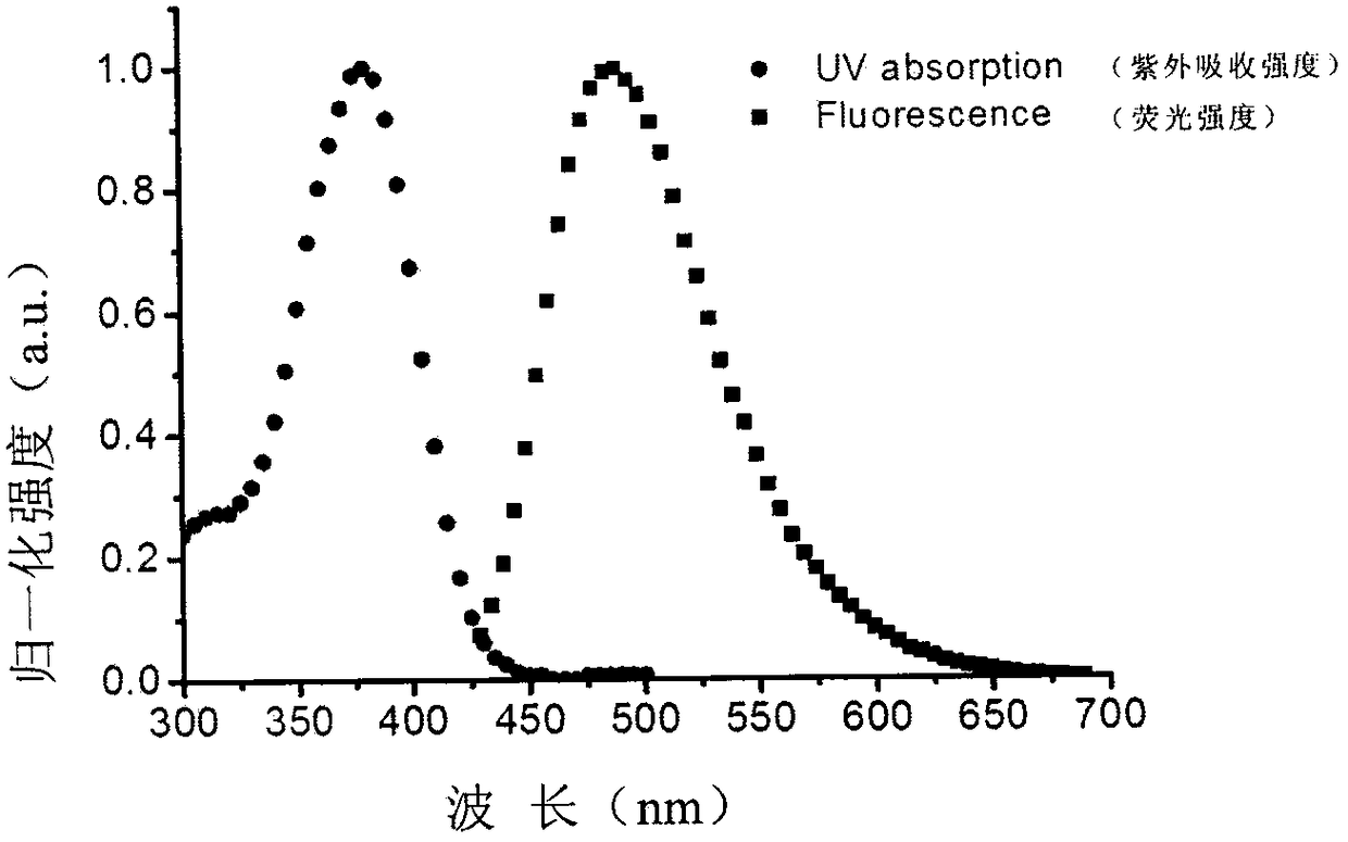
[0060] Prepare a TAB-3-cRGD probe PBS solution with a concentration of 10 μM, add 3 mL to a 10mm*10mm two-way cuvette, put it into a spectrophotometer with a baseline adjusted, and test the data in the range of 500-300; then Pour the solution into a 10mm*10mm four-way cuvette, use 405nm as the excitation wavelength, and test the fluorescence spectrum in the range of 430-700nm (see figure 1 ). Depend on figure 1 It can be seen that the maximum absorption peak of TAB-3-cRGD is at 380nm, and the maximum emission peak is at 490nm.
Embodiment 3
[0061] Example 3: Imaging application of TAB-3-cRGD fluorescent probe in living cells
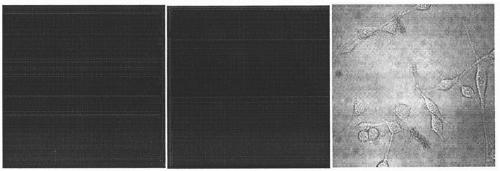
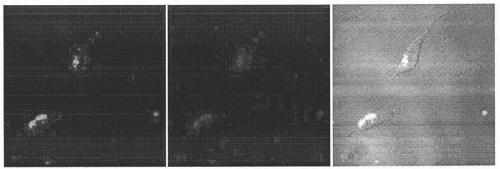
[0062] Mouse fibroblasts NIH / 3T3, venous endothelial cells HUVEC-1, and glioma U87 cells were cultured in a confocal culture medium (the volume ratio of DMEM medium and fetal bovine serum in the medium was 9:1). on the dish. Place them in an incubator at 37° C., 5% (volume fraction) CO2 and 20% (volume fraction) O2, and cultivate them for 24-48 hours. Remove the culture medium, wash it three times with PBS, then add serum-free DMEM culture solution, add the solution of the TAB-3-cRGD fluorescent probe of the present invention in each culture dish respectively, continue to cultivate in the incubator for 15min, PBS ( Phosphate buffered saline) to wash the cultured cells 6 times. Fluorescent imaging was performed using a confocal microscope. The imaging conditions are: the excitation wavelength is 405nm, and the collection range is: 500-550nm. Such as figure 2 is the imaging image of TAB...
PUM
 Abstract
Description
Claims
Application Information
 - R&D Engineer
- R&D Manager
- IP Professional
- Industry Leading Data Capabilities
- Powerful AI technology
- Patent DNA Extraction
Browse by: Latest US Patents, China's latest patents, Technical Efficacy Thesaurus, Application Domain, Technology Topic, Popular Technical Reports.
© 2024 PatSnap. All rights reserved.Legal|Privacy policy|Modern Slavery Act Transparency Statement|Sitemap|About US| Contact US: help@patsnap.com


