
Production method of skin pathological section

A production method and technology of pathological slices, applied in the biological field, can solve the problems of waste specimens, incomplete information of pathological pictures, poor consistency between pathological specimens and living tissues, etc., and achieve the effect of avoiding shrinkage

Inactive Publication Date: 2018-08-03
THE FIRST AFFILIATED HOSPITAL OF SUN YAT SEN UNIV
View PDF2 Cites 3 Cited by
  • Summary
  • Abstract
  • Description
  • Claims
  • Application Information

AI Technical Summary

Problems solved by technology

[0003] In order to solve the above problems, the present invention provides a method for making skin pathological slices, which standardizes the methods of taking, fixing and embedding skin tissues, and solves the problems of poor consistency between pathological specimens and living tissues, incomplete information of pathological pictures, and waste of specimens. It can also provide self-control for skin tissue immunohistochemistry and immunofluorescence staining without significantly increasing the difficulty of operation.

Method used


Image

  • Production method of skin pathological section
  • Production method of skin pathological section
  • Production method of skin pathological section

Examples


Embodiment 1

[0025] A method for making a skin pathology slice, comprising the steps of:

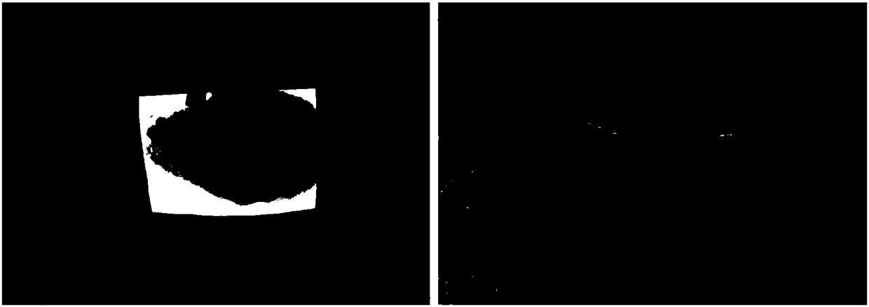
[0026] (1) Rat skin tissue collection: spread a heating blanket and white paper on the experimental table for dead rats, place the rats in a prone position, take a blue surgical drape, and cut a square opening in the middle for To expose the surgical field of view, align the opening on the back of the rat and lay it on top of the rat. The central axis of the opening of the surgical drape is consistent with the midline of the back of the rat; For complete hair removal in the target area, place the transparent film on the target area and use an oil-based pen to trace and record the size and shape of the target area, see figure 1 ;Take another small piece of paper to record the experiment time and subjects, and use ophthalmic scissors to cut the skin of the target area on the back of the rat, down to the muscle level; after removing the tissue, the epidermis, dermis and subcutaneous tissue can be seen f...


PUM

PropertyMeasurementUnit
thicknessaaaaaaaaaa

Abstract

The invention discloses a production method of a skin pathological section. The method comprises the following steps: obtaining a material from a living body and fixing in-vitro skin tissues; dehydrating in-vitro tissues, immersing with wax, embedding, sliding and dyeing to prepare the skin pathological section. Material taking, fixing and embedding methods of animal skin and subcutaneous tissuesare standardized and the problems that the consistency of a skin pathological specimen and a living body tissue shape is poor and information of a pathological picture is not complete are solved; under the condition that the operation difficulty is not remarkably improved, self control also can be provided for skin immunohistochemistry and immunofluorescence staining.

Description

technical field [0001] The invention relates to the field of biotechnology, in particular to a method for making pathological skin slices. Background technique [0002] Skin tissue is highly elastic, complex in structure, and multi-layered. Different species and sources of skin have different levels of connection and density between layers of tissue. Skin tissue is roughly divided into complex structures such as epidermis, dermis, subcutaneous tissue, and skin appendages. , the density of these tissues is not the same, the stratum corneum is relatively denser, and the skin tissue is prone to curling and deformation when it is processed by conventional sampling and fixing methods, not only cannot restore the original shape of the living tissue, but also often encounters only The problem of obtaining oblique sections of epidermis / dermis / subcutaneous tissue instead of full-thickness vertical sections of skin will not only cause waste of experimental specimens, but also cause la...

Claims


Application Information

Patent Timeline
Patent Type & Authority Applications(China)
IPC IPC(8): G01N1/28G01N1/30G01N1/36G01N1/06G01N1/04
CPCG01N1/04G01N1/06G01N1/286G01N1/30G01N1/36G01N2001/2873
Inventor 陈蕾周飞祁少海吴燕茹王凯悦孙秀红
Owner THE FIRST AFFILIATED HOSPITAL OF SUN YAT SEN UNIV
Learn More