Detection method of fish allergen
A detection method and technology for allergens, applied in the field of food analysis, can solve the problems of low sensitivity and precision, complicated operation, long detection time, etc., and achieve the effects of accurate results, strong specificity and easy operation.
- Summary
- Abstract
- Description
- Claims
- Application Information
AI Technical Summary
Problems solved by technology
Method used
Image
Examples
Embodiment 1
[0061] The preparation of embodiment 1 sample
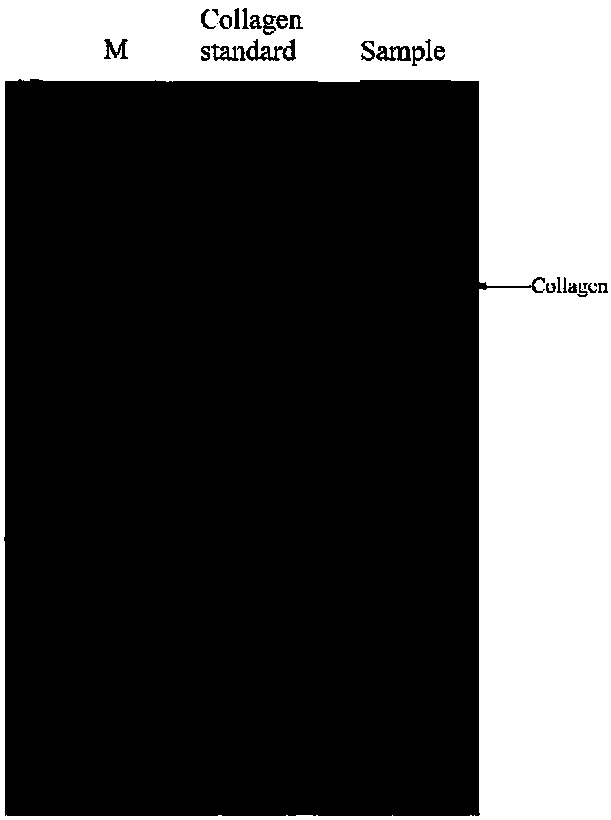
[0062] Sample preparation: take 2g of tuna flesh, grind it into powder with liquid nitrogen, and then use the KGI Complete Protein Extraction Kit to extract the protein infusion. The specific steps are as follows: (1) Add 1 μL of protease inhibitor, 10 μL of phosphatase inhibitor and 5 μL of 100 mM PMSF to 1 mL of pre-cooled Lysis Buffer, mix well and store on ice for several minutes before use; (2) Take fresh tuna , Cut the fish meat into mud with a knife, put it in a mortar, add liquid nitrogen and grind it until it is powdery. (3) Quickly weigh 0.1g of fish meal and put it in a 1.5mL pre-cooled centrifuge tube, add 1mL of the above mixture, mix well and let stand at 4°C for 2h; (4) Centrifuge at 10000r / min for 5min at 4°C, take the supernatant , aliquoted and stored at -80°C, repeated freezing and thawing should be avoided. The sample solution was analyzed by SDS-PAGE electrophoresis, and the results were as follows figure...
Embodiment 2
[0063] Example 2 Coating of Optical Fiber and Collagen Antigen
[0064] First use piraha solution (H 2 SO 4 :H 2 o 2 =3:1) Soak the optical fiber probe for 30 minutes, fully clean it with ultrapure water, and blow it dry with nitrogen gas to make the surface of the probe hydroxylated; Next, soak the optical fiber probe with 5% (v / v) glutaraldehyde solution at 37°C for 2 hours, wash it thoroughly with toluene, and dry it in an oven at 120°C to silanize the surface of the probe; put the treated optical fiber probe into 2.5mg / mL collagen solution, soak overnight at 4°C to connect the coated antigen collagen; rinse the fiber optic probe with ultrapure water, and then soak it in 4mg / mL BSA solution for 2h to seal the non-specific adsorption point. The coated fiber can be stored at 4°C for several months.
Embodiment 3
[0065] Example 3Cy5.5 Fluorescent Dye Labeled Collagen Monoclonal Antibody (Collagen Monoclonal Antibody)
[0066] Dialyze the collagen monoclonal antibody solution with 0.15M NaCl solution at room temperature for 4 hours, change the solution, dialyze overnight at 4°C, and then dialyze with 0.1M NaHCO 3 (pH 8.3) solution was dialyzed at room temperature for 4 h, then filtered the collagen monoclonal antibody solution (3 mg / mL) with a 0.22 μm syringe filter head, and added Cy5.5 solution ( 10mg / mL), protected from light, stirred slowly for 40min to fully combine the antibody with Cy5.5, and then dialyzed the Cy5.5-labeled collagen monoclonal antibody solution with NaCl solution at room temperature in the dark for 4h to remove unlabeled Cy5. 5. Change the medium and dialyze overnight at 4°C in the dark, then dialyze the Cy5.5-labeled collagen monoclonal antibody in PBS+0.01% sodium azide solution at room temperature in the dark for 4 hours, change the medium, and dialyze overnig...
PUM
| Property | Measurement | Unit |
|---|---|---|
| concentration | aaaaa | aaaaa |
| concentration | aaaaa | aaaaa |
Abstract
Description
Claims
Application Information
 - Generate Ideas
- Intellectual Property
- Life Sciences
- Materials
- Tech Scout
- Unparalleled Data Quality
- Higher Quality Content
- 60% Fewer Hallucinations
Browse by: Latest US Patents, China's latest patents, Technical Efficacy Thesaurus, Application Domain, Technology Topic, Popular Technical Reports.
© 2025 PatSnap. All rights reserved.Legal|Privacy policy|Modern Slavery Act Transparency Statement|Sitemap|About US| Contact US: help@patsnap.com


