Kit for detecting markers of ovarian cancer cells in peripheral blood
An ovarian cancer cell and kit technology, which is applied in the biological field, can solve the problems of being unable to be used as an indicator for detecting tumor progression and being highly invasive, and achieve the effects of less damage, small sample size and high detection sensitivity.
- Summary
- Abstract
- Description
- Claims
- Application Information
AI Technical Summary
Problems solved by technology
Method used
Image
Examples
Embodiment 1
[0080] Example 1: Preparation of magnetic beads bound to ovarian cancer antibody:
[0081] Antibodies used were anti-EpCAM monoclonal antibody, anti-Muc1 monoclonal antibody and anti-Her-2 monoclonal antibody. This kit uses the above three antibodies to be labeled separately, and then they are mixed for use.
[0082] Labeling method: magnetic beads produced by Invitrogen, USA M-450Tosylactivated. The marking method is strictly in accordance with the product instructions.
[0083] The labeling ratio is: label 500 μL magnetic beads per 100 μg antibody, and mix in equal amounts after labeling.
Embodiment 2
[0084] Example 2: Isolation of ovarian cancer cells:
[0085] 2.1 Sample handling:
[0086] Take 5mL of peripheral blood from patients with ovarian cancer, anticoagulate with EDTA, store at 4 degrees, and use within 48 hours.
[0087] 2.2 Magnetic bead treatment:
[0088] Wash the magnetic beads bound with ovarian cancer antibody (antibody-labeled magnetic beads, which are mixed magnetic beads labeled with three antibodies, and prepare antibody-labeled magnetic beads according to the ratio of 1 mL sample to 25 μL magnetic beads) with PBS repeatedly for three times, and separate the magnetic beads. Beads, kept on ice;
[0089] The specific operation is: draw 125μL of marked magnetic beads into a 1.5mL centrifuge tube, gently blow and mix with a pipette (note that a vortexer cannot be used), and then place it on the magnetic bead enricher for 1min. , to attach the magnetic beads to the wall of the centrifuge tube, discard the supernatant; then add 1mL PBS, place it on the mag...
Embodiment 3
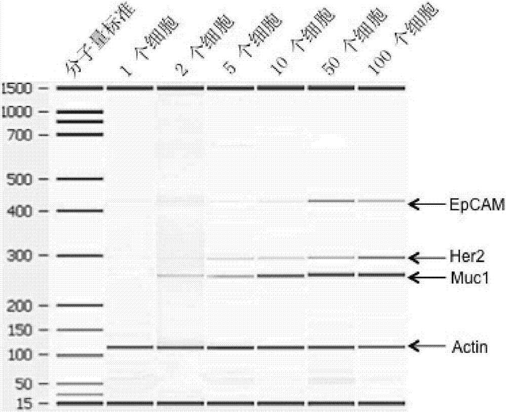
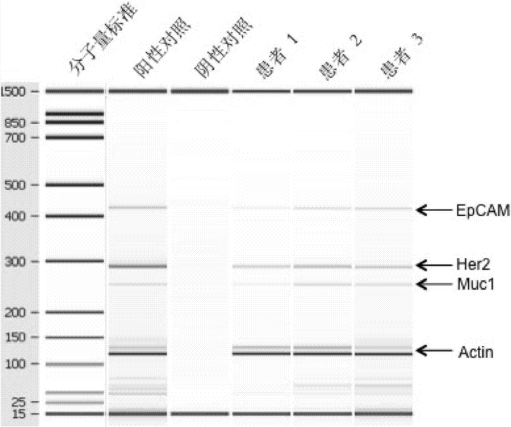
[0096] Example 3: Detection of ovarian cancer cell markers:
[0097]3.1 mRNA purification
[0098] Mix 40 μL magnetic beads with oligonucleotide Oligo(dT) into a 1.5 mL centrifuge tube, add 500 μL lysis / binding buffer to wash twice, separate magnetic beads, add supernatant, mix well, and incubate at room temperature 10min, let the mRNA bind to the magnetic beads; then separate the magnetic beads, wash the magnetic beads twice with 500 μL buffer A, separate the magnetic beads, then wash the magnetic beads twice with 500 μL buffer B, separate the magnetic beads, and then use 100 μL RNase-free Wash once with water, separate the magnetic beads, resuspend the magnetic beads with 29.5 μL RNase-free water, incubate in a water bath (or metal bath) at 55°C for 5 min, and place on ice for 2 min to obtain the mRNA / magnetic bead mixture.
[0099] The mRNA / bead mix cannot be stored and needs to be reverse transcribed immediately.
[0100] 3.2 Reverse transcription:
[0101] The reverse ...
PUM
 Abstract
Description
Claims
Application Information
 - R&D
- Intellectual Property
- Life Sciences
- Materials
- Tech Scout
- Unparalleled Data Quality
- Higher Quality Content
- 60% Fewer Hallucinations
Browse by: Latest US Patents, China's latest patents, Technical Efficacy Thesaurus, Application Domain, Technology Topic, Popular Technical Reports.
© 2025 PatSnap. All rights reserved.Legal|Privacy policy|Modern Slavery Act Transparency Statement|Sitemap|About US| Contact US: help@patsnap.com


