
Method for detecting enzyme digestion of extracellular fragment of platelet receptor GPIba based on flow cytometry

A technology of flow cytometry and platelets, applied in the field of medical biology, can solve the problems of high background value of ELISA, lower reliability and stability of test results, unsuitable detection of GPIba extracellular segment enzyme digestion, etc., to achieve good stability, The effect of high reliability

Active Publication Date: 2017-11-10
XUZHOU MEDICAL UNIV
View PDF6 Cites 1 Cited by
  • Summary
  • Abstract
  • Description
  • Claims
  • Application Information

AI Technical Summary

Problems solved by technology

However, Western blot detection requires a certain number of cells to obtain the amount of protein required for detection, so in the case of thrombocytopenia, this method is not suitable for detection of GPIba extracellular segment enzyme digestion
In addition, cleavage of the extracellular segment of GPIba occurs continuously (even at rest), which can lead to high levels of soluble fragments in plasma, serum, or platelet supernatants, resulting in high background values ​​in ELISA
Detecting GPIba extracellular segment digestion by ELISA method will reduce the reliability and stability of the detection results

Method used


Image

  • Method for detecting enzyme digestion of extracellular fragment of platelet receptor GPIba based on flow cytometry
  • Method for detecting enzyme digestion of extracellular fragment of platelet receptor GPIba based on flow cytometry
  • Method for detecting enzyme digestion of extracellular fragment of platelet receptor GPIba based on flow cytometry

Examples


Embodiment 1

[0040] Example 1: Detection of enzyme cleavage of GPIba extracellular segment of healthy human platelets

[0041] A method for detecting the cleavage of the extracellular segment of platelet receptor GPIba based on flow cytometry, comprising the following steps:

[0042] (1) Sample preparation and processing

[0043] a. Collect 5ml of venous blood from healthy people, and collect in a test tube containing an aqueous solution of anticoagulant trisodium citrate (1.6 grams of trisodium citrate dissolved in 50ml of distilled water), the aqueous solution of anticoagulant trisodium citrate and intravenous The blood is mixed slowly and gently according to the volume ratio of 1:9;

[0044] b. Centrifuge at 120×g for 20 minutes at room temperature, and use a plastic pipette to gently suck up the platelet-rich plasma (PRP) in the upper layer. Do not suck up the PRP near the middle layer, so as not to suck white blood cells and cause pollution;

[0045] c. Add the sucked PRP to 5 EP tu...

Embodiment 2

[0061] Example 2: Detection of NEM-treated healthy human platelets GPIba extracellular fragmentation

[0062] N-ethylmaleimide (NEM), a reagent that can artificially induce the cleavage of the extracellular segment of GPIba, was used to treat platelets and induce cleavage of GPIba.

[0063] from Figure 7 It can be seen that the abscissa is the binding of intracellular segment antibodies, and the ordinate is the binding of extracellular segment antibodies. The four boxes in the figure represent the binding of each antibody, respectively. The lower left quadrant represents the negative area, i.e. what percentage of platelets have neither extracellular nor intracellular antibody binding. The upper left quadrant represents what percentage of platelets have only extracellular antibody bound. The lower right quadrant represents platelets with intracellular segment antibodies bound. The upper right quadrant represents the percentage of platelets that contain both extracellular a...

Embodiment 3

[0066] Example 3: Detection of platelet GPIba extracellular fragmentation in patients with thrombocytopenia

[0067] The platelets of 5 patients with thrombocytopenia were collected, and the method of the present invention was used to detect GPIba extracellular segment enzyme cleavage. from Figure 9 It can be seen that the intact state of GPIba was significantly reduced in patients with thrombocytopenia, suggesting an increase in extracellular fragment cleavage. In addition, through correlation analysis, it was concluded that the degree of cleavage of the extracellular segment of GPIba in patients with thrombocytopenia did not depend on changes in the number of platelets (R2=0.021, P=0.815), which further verified the flow cytometry-based detection of the present invention. The method of cleavage of the extracellular segment of GPIba does not depend on changes in platelet number.


PUM


Abstract

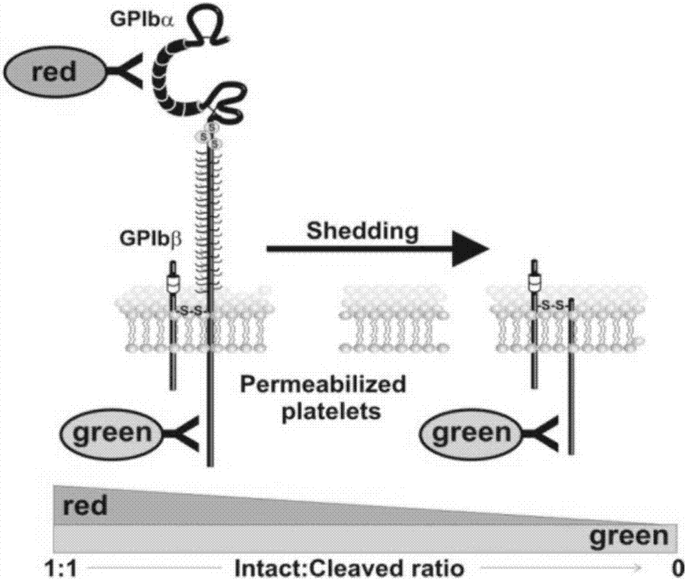
The invention provides a method for detecting enzyme digestion of the extracellular fragment of a platelet receptor GPIba based on flow cytometry. The method comprises the following concrete steps: (1) preparation and treatment of samples; (2) detection with a flow cytometer; and (3) data analysis with Weasel software. According to the invention, two different fluorescently-labeled antibodies are used, i.e., an anti-GPIba extracellular fragment antibody and an anti-GPIba intracellular fragment antibody; and the relative value of bonding of the extracellular fragment antibody and the intracellular fragment antibody is analyzed by using flow cytometry, so the degree of enzyme digestion of the GPIba extracellular fragment can be reflected. The detection method provided by the invention is independent on the changes of the number of platelets and applicable to the situation of reduction in the number of platelets, and has high reliability and good stability.

Description

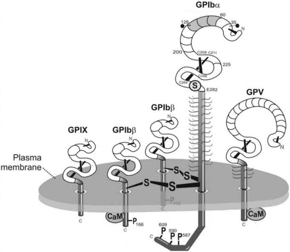
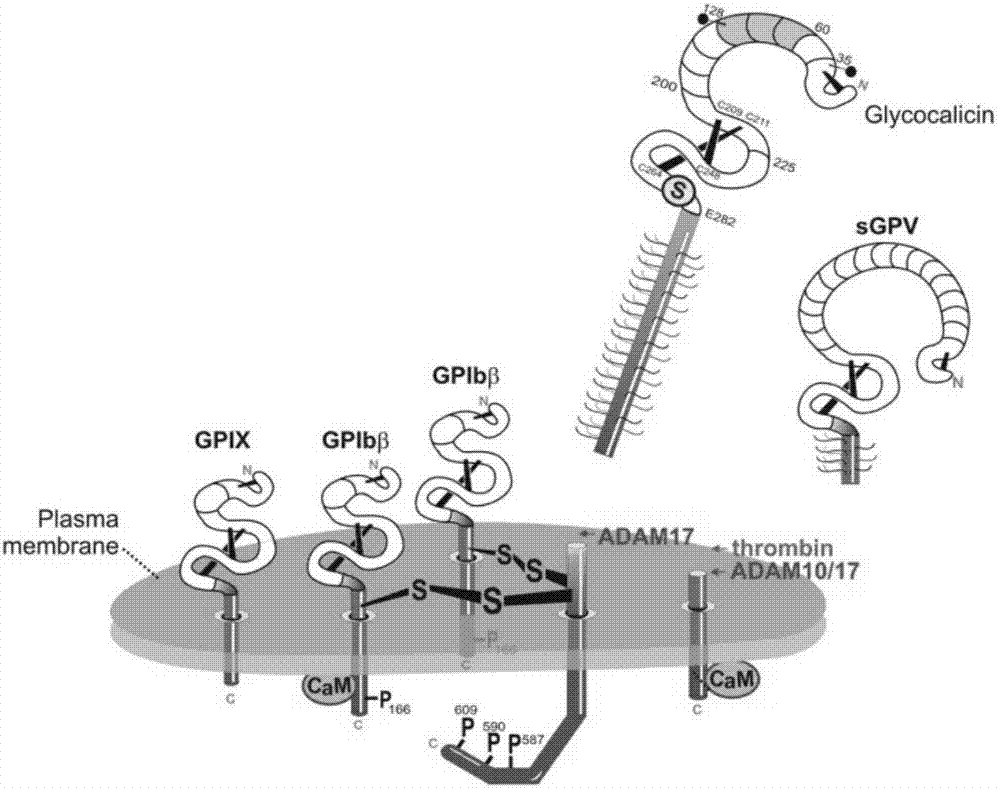
technical field [0001] The invention belongs to the technical field of medical biology, and in particular relates to a method for detecting the enzyme cleavage of the extracellular segment of platelet receptor GPIba based on flow cytometry. Background technique [0002] The GPIb-IX-V complex consists of GPIba, GPIbb, GPIX and GPV ( figure 1 ). GPIba binds to GPIbb through disulfide bonds, and then connects to GPIX and GPV through non-covalent bonds. GPIba is the major ligand-binding subunit in the GPIb-IX-V complex and consists of 626 amino acid residues. GPIba is a membrane-expressed molecule consisting of an extracellular segment, a transmembrane region and an intracellular segment. As the most important adhesion molecule of platelets, GPIba exerts various biological functions by binding with corresponding ligands. [0003] After platelet activation or some kind of stimulation, the extracellular segment of GPIba will undergo enzymatic cleavage, which is mainly mediated...

Claims


Application Information

Patent Timeline
Patent Type & Authority Applications(China)
IPC IPC(8): G01N15/14G01N1/28G01N1/30G01N33/58
CPCG01N1/28G01N1/30G01N15/14G01N33/582
Inventor 乔建林徐开林曾令宇鞠文齐昆明
Owner XUZHOU MEDICAL UNIV
Learn More