3D printing biological bracket with drug release function and preparation method thereof

A 3D printing and biological stent technology, applied in pharmaceutical formulations, drug delivery, pharmaceutical sciences, etc., can solve the problems of difficult drug release, difficult combination of multiple drugs, and difficult loading of drugs.

Active Publication Date: 2017-06-30
SHANGHAI UNION TECH
View PDF5 Cites 6 Cited by
  • Summary
  • Abstract
  • Description
  • Claims
  • Application Information

AI Technical Summary

Problems solved by technology

However, the existing 3D printed scaffolds only satisfy the structure, and it is difficult to meet the biological function requirements of promoting regeneration.
Drugs have the functions of treating diseases and promoting regeneration. However, due to the influence of the preparation process, it is difficult to realize the controlled release of drugs in the stent, especially the release of drugs such as active macromolecules and active factors.
In the existing technology, the drug release function of the stent is mainly realized by coating the drug on the surface of the stent, but it is difficult to load the drug inside the stent, and it is difficult to realize the sustained release of the drug, and it is difficult to realize the loading of proteins, water-soluble drugs, etc., especially protein The activity of similar drugs is maintained, and it is difficult to realize the combined effect of multiple drugs

Method used


Image

  • 3D printing biological bracket with drug release function and preparation method thereof
  • 3D printing biological bracket with drug release function and preparation method thereof
  • 3D printing biological bracket with drug release function and preparation method thereof

Examples


Embodiment 1

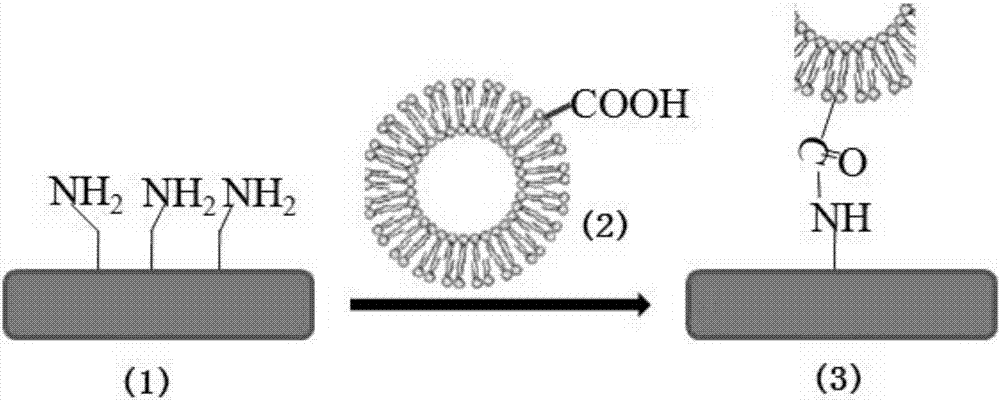
[0042] After mixing 1 g of PLLA and 0.13 g of KH550 (aminopropyltriethoxysilane), 3D printing was carried out to obtain a 3D printed scaffold with surface amination.

[0043] According to the molar ratio of 96:19:4:20, weigh dipalmitoylphosphatidylcholine, cholesterol, DSPE-PEG-COOH (phosphatidylethanolamine-polyethylene glycol-carboxy) and paclitaxel, and dissolve it with 30mL chloroform Transfer to an eggplant-shaped bottle. The lipid film was obtained after the solvent was evaporated by rotary evaporation, and dried in vacuum overnight. Add 20 mL of PBS to the eggplant-shaped bottle for hydration to obtain liposome crude emulsion. The liposome coarse emulsion was processed with an ultrasonic probe, the ultrasonic time was 3 minutes, the ultrasonic power was 40%, and the operation was 2 seconds and the stop was 1 second. The sonicated liposome solution is sequentially passed through 0.8 μm, 0.45 μm, and 0.22 μm microporous membranes to obtain a liposome solution modified w...

Embodiment 2

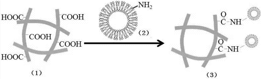
[0047] The cleaned and dried 3D printed scaffold (made of PLLA) was soaked in 6wt% hexamethylenediamine / n-propanol solution and shaken at 60°C for a certain period of time, then washed repeatedly with ethanol and deionized water and dried in vacuum to obtain amino-functionalized scaffolds. 3D printed brackets.

[0048] According to the molar ratio of 80:40:4:20, weigh soybean lecithin, cholesterol sulfate, DSPE-PEG-COOH (phosphatidylethanolamine-polyethylene glycol-carboxy) and ibuprofen, then dissolve them with 30mL chloroform and transfer into an eggplant-shaped bottle. The lipid film was obtained after the solvent was removed by rotary evaporation, and the organic residue was removed by vacuum drying overnight. Add 20 mL of PBS to the eggplant-shaped bottle for hydration to obtain liposome crude emulsion. The liposome coarse emulsion was processed with an ultrasonic probe, the ultrasonic time was 3 min, the ultrasonic power was 20%, and the working time was 1 second and t...

Embodiment 3

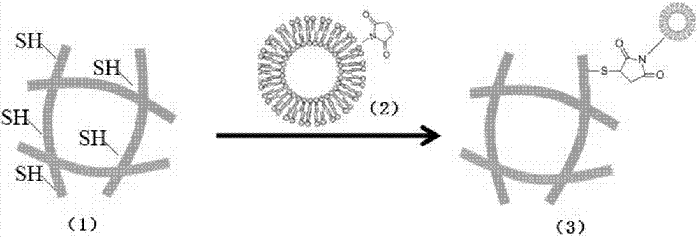
[0051] Using 3D printing technology to print out the PCL bioscaffold. Plasma treatment was performed on the bracket, as follows: the 3D printed bracket was placed in the plasma device and fixed between the electrodes, and when the pressure in the inner chamber reached 10 -3 At Torr, inject oxygen and gaseous acrylic acid at 0.2 Torr, and apply radio frequency power of 50W and negative electrode pulse voltage for 30s to obtain a 3D printed scaffold with carboxyl groups on the surface.

[0052] According to the molar ratio of 90:30:3:15, weigh egg yolk lecithin, cholesterol, DSPE-PEG-NH 2 (phosphatidylethanolamine-polyethylene glycol-amino) and doxorubicin, and dissolve it in 2mL ethanol solution to obtain a lipid ethanol solution. Measure 20 mL of PBS solution and place it in a beaker, and add the lipid ethanol solution dropwise into the beaker under the conditions of a rotating speed of 300 rpm and a temperature of 40°C. After the dropwise addition is completed, continue to ...


PUM


Abstract

The invention relates to a preparation method of a 3D printing biological bracket with a drug release function. The method comprises the following steps: providing a 3D printing biological bracket with active groups being modified on the surface; providing drug-carrying liposome solution with active groups being modified on the surface; immersing the biological bracket into the drug-carrying liposome solution at 4 to 40 DEG C, and enabling the active groups modified on the surfaces of the printing biological bracket and the drug-carrying liposome solution to react, so as to obtain a 3D printing biological bracket with a drug release function. The invention further provides a 3D printing biological bracket with a drug release function prepared through the preparation method. A number of special groups are modified on the surface of the biological bracket, a number of special groups, such as carboxyl or amidogen, are grafted on the surface of a liposome bimolecular layer, then functional groups on the surfaces of the printing biological bracket and the drug-carrying liposome solution are enabled to react, the nanometer liposome is combined on the surface of the biological bracket, and loading of different drugs is realized through liposome, so that the biological bracket with conventional functions has a treating function.

Description

technical field [0001] The invention relates to the technical field of preparation of biological scaffolds, in particular to a 3D printed biological scaffold with drug release function and a preparation method thereof. Background technique [0002] Implantable medical bioscaffolds have the advantages of structural bionics and tissue plasticity, and bioscaffolds based on 3D printing technology have been widely used in the medical field. At present, implantable bioscaffolds mainly play the role of structural biomimesis. Implantable scaffolds not only need to have the effect of structural reconstruction, but also need excellent biocompatibility, so as to improve the interfacial bonding force between the scaffold and the tissue. [0003] At present, the materials of these biological scaffolds are mainly metals and polymers. 3D printing mainly focuses on structural design to meet the special functions of 3D structures. However, for 3D scaffolds implanted in vivo to repair tissu...

Claims


Application Information

Patent Timeline
IPC IPC(8): A61L27/18A61L27/34A61L27/54
CPCA61L27/18A61L27/34A61L27/54A61L2300/602A61L2300/626
Inventor 崔文国余嘉程若昱许建辉杨林
Owner SHANGHAI UNION TECH
Why Eureka
Try Eureka