Human pituitary adenoma cell line and application thereof
A technology for pituitary adenomas and uses, applied in the field of tumor biology, can solve the problems of pituitary tumor cells without humanization, difficult to transfer, etc.
- Summary
- Abstract
- Description
- Claims
- Application Information
AI Technical Summary
Problems solved by technology
Method used
Image
Examples
Embodiment 1
[0040] Embodiment 1: Acquisition and cultivation of human pituitary adenoma cell line
[0041] 1. HPA1446 primary cell culture
[0042] 1. Source of biological material
[0043] The cell line of the present invention is obtained by separating and culturing the pituitary tumor tissue of a non-functioning pituitary tumor patient. The tumor tissue was obtained from a surgical specimen of a patient in the Neurosurgery Center of Beijing Tiantan Hospital Affiliated to Capital Medical University. The pathological diagnosis was a non-functioning pituitary adenoma. Under the microscope, the tumor cells were densely distributed, the boundaries of the cell membrane were unclear, and the size and shape of the nuclei were not consistent. , nucleoli and binuclear tumor cells can be seen.
[0044] 2. Isolation and culture method
[0045] Wash the tissue block twice with PBS solution (1:1) containing red solution (purchased from Gibco) in a sterile ultra-clean bench, remove blood vessels a...
Embodiment 2
[0064] Embodiment 2: Determination of cell growth curve
[0065] 1. Method:
[0066] The primary pituitary adenoma cells of Example 1 and the immortalized pituitary adenoma cells were adjusted to a cell concentration of 5×10 cells after the cells were overgrown. 4 cells / mL, inoculate primary pituitary tumor cells and immortalized pituitary tumor cells in 96-well culture plate, 100 μl per well, 37°C, 5% CO 2 Cultivate in an incubator; add 10 μl of MTT to each well after culturing for 12h, 24h, 36h, 48h, and 72h respectively, at 37°C, 5% CO 2 Cultivate in the incubator and incubate for 3-4 hours in the dark; absorb the liquid in the well, add 200 μl of DMSO, and shake on the shaker at room temperature for 10 minutes; measure the OD value at the same time point with a wavelength of 492nm on a microplate reader, and use the measured OD value analyze.
[0067] 2. Results:
[0068] Such as figure 1 As shown, the immortalized pituitary tumor cells entered the growth stationary p...
Embodiment 3
[0069] Embodiment 3: cell morphology detection
[0070] 1. Method:
[0071] 1. Observe the cell line CGMCC No.12672 with a phase contrast microscope.
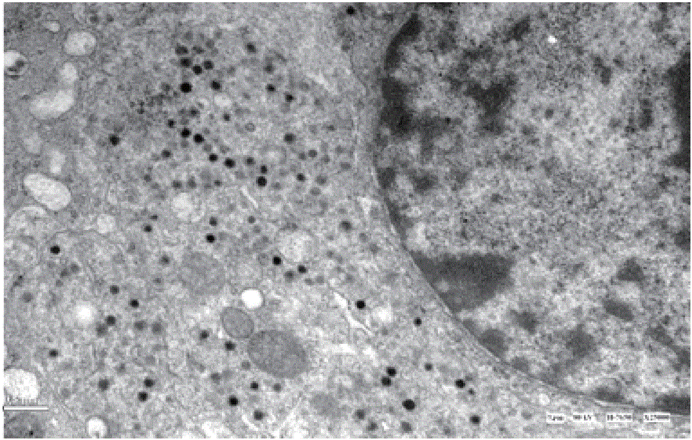
[0072] 2. Observing the ultrastructure of the cell line CGMCC No.12672 with a transmission electron microscope:
[0073] When the cells grow to 90% confluence, discard the culture medium, add 3ml 0.1MPBS (pH value 7.4), scrape the cells with a rubber cell scraper, collect the cells, centrifuge (1000rpm / min, 10 minutes), discard the supernatant, Aldehyde / osmium tetroxide pre-fixed for 2 hours, washed 3 times with 0.1M PBS (pH 7.4), 10 minutes each time. Then the specimens were post-fixed, dehydrated, soaked, embedded, cut into ultra-thin sections and stained with heavy metals (completed by the electron microscope room of Beijing Institute of Neurosurgery).
[0074] 2. Results:
[0075] 1. The growth characteristics of the cell line CGMCC No.12672 under the phase-contrast microscope: most of them are fusiform, some are triang...
PUM
 Abstract
Description
Claims
Application Information
 - R&D
- Intellectual Property
- Life Sciences
- Materials
- Tech Scout
- Unparalleled Data Quality
- Higher Quality Content
- 60% Fewer Hallucinations
Browse by: Latest US Patents, China's latest patents, Technical Efficacy Thesaurus, Application Domain, Technology Topic, Popular Technical Reports.
© 2025 PatSnap. All rights reserved.Legal|Privacy policy|Modern Slavery Act Transparency Statement|Sitemap|About US| Contact US: help@patsnap.com


