A method and magnetic resonance imaging system for monitoring the temperature of tissue surrounding an active implant

A magnetic resonance imaging and surrounding tissue technology, applied in the field of medical equipment, can solve problems such as MRI signal loss, surrounding magnetic field distortion, and inability to obtain temperature information, and achieve the effect of eliminating potential safety hazards

Active Publication Date: 2018-11-23
TSINGHUA UNIV
View PDF7 Cites 0 Cited by
  • Summary
  • Abstract
  • Description
  • Claims
  • Application Information

AI Technical Summary

Problems solved by technology

MR thermometry is only used as a monitoring method
For the radiofrequency heating of implantable medical devices under nuclear magnetic resonance, the heating comes from the MRI scan itself, and the scanning sequences have their own purposes and have different parameters. They are very different from the scanning of MR thermometry, and cannot be obtained from scanning sequences for other purposes. Get temperature information from
[0014] In addition, metal conductors in implantable medical devices will also cause distortion of the surrounding magnetic field due to magnetization under the magnetic field of MRI, which will cause image artifacts, making the MRI signal near the conductor lost or seriously distorted
The radio frequency induction heating is the most serious near the conductor, which is also a problem in the current application of MR thermometry.

Method used


Image

  • A method and magnetic resonance imaging system for monitoring the temperature of tissue surrounding an active implant
  • A method and magnetic resonance imaging system for monitoring the temperature of tissue surrounding an active implant
  • A method and magnetic resonance imaging system for monitoring the temperature of tissue surrounding an active implant

Examples


Embodiment Construction

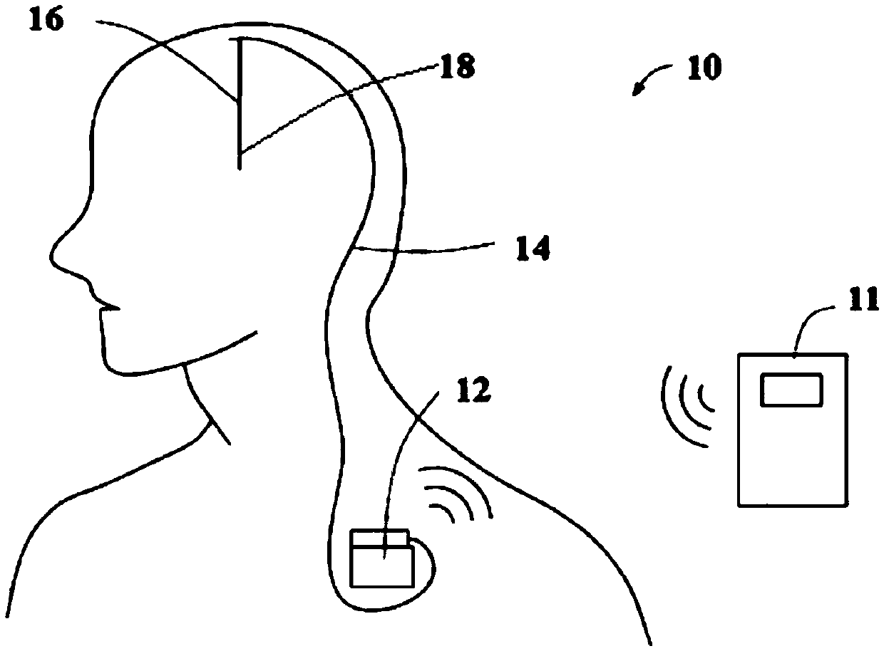
[0058] The invention provides a method for real-time monitoring of tissue temperature around an active implant under MR based on MR temperature measurement and a safety assessment and a magnetic resonance imaging system using the method. The active implant can be a cardiac pacemaker, defibrillator, deep brain stimulator, spinal cord stimulator, vagus nerve stimulator, gastrointestinal stimulator or other similar implanted medical devices. The present invention is only described by taking the deep brain electrical stimulator as an example, and the present invention is further described in conjunction with the accompanying drawings.

[0059] See figure 1 , the deep brain electrical stimulator 10 includes: an external programmer 11, a pulse generator 12 implanted in the body, an extension wire 14 and a stimulating electrode 16. The external programmer 11 controls the pulse generator 12 to generate a certain pattern of current pulses, which are transmitted to the electrode contac...


PUM


Abstract

A method for monitoring the temperature of tissues surrounding an active implant, the method being based on the magnetic resonance temperature detection technology and using a magnetic resonance imaging (MRI) system (20); the MRI system (20) at least comprising a sequence 2 for clinical examination or scientific research or other uses and a sequence 3 for measuring temperature distribution. The method comprises the following steps: step S11, performing scan using a sequence 2, and performing the scan of a temperature measurement sequence 3 alternately in the sequence 2; and step S12, performing a safety assessment according to the scanning result of the temperature measurement sequence 3. The method can effectively monitor the radio-frequency temperature rise of a patient wearing an implantable medical device who is undergoing an MRI scan, removing potential safety risk.

Description

technical field [0001] The present invention relates to the technical field related to medical devices, in particular, to a method for real-time monitoring of tissue temperature around active implants under MR based on magnetic resonance (Magnetic Resonance, MR) temperature measurement technology and a magnetic resonance imaging system using the method . Background technique [0002] Compared with other imaging techniques (such as X-ray, CT, etc.), Magnetic Resonance Imaging (MRI) has significant advantages: MRI is clearer, has a high resolution for soft tissues, and There is no ionizing radiation damage to the human body. Therefore, magnetic resonance imaging technology is widely used in the clinical diagnosis of modern medicine. Today, it is estimated that at least 60 million cases worldwide are examined using MRI technology every year. [0003] When MRI works, three magnetic fields come into play. A high-intensity uniform static magnetic field B 0 , a gradient field ...

Claims


Application Information

Patent Timeline
Patent Type & Authority Patents(China)
IPC IPC(8): A61B5/055A61B5/01
CPCA61B5/01A61B5/055
Inventor 姜长青张锋丁建琦董延涛李路明
Owner TSINGHUA UNIV
Why Eureka
Try Eureka