
Radioactive C-MET-targeted affinity micromolecular compound and application thereof

A small molecule compound, radioactive technology, applied in the directions of radioactive carriers, radioactive preparations in vivo, introduction of heterocyclic compound isotopes, etc., can solve the problems of inability to realize dynamic research, unfavorable dynamic research, complicated operation, etc., and achieve good biological distribution characteristics, The effect of good pharmacokinetic characteristics and accurate test results

Active Publication Date: 2016-10-12
上海准视生物科技有限公司
View PDF2 Cites 7 Cited by
  • Summary
  • Abstract
  • Description
  • Claims
  • Application Information

AI Technical Summary

Problems solved by technology

Although in vitro detection methods can judge and analyze the expression level of C-MET in cells or tissues, there are certain limitations in terms of technical analysis: ① Specimens need to be obtained through cell culture, biopsy or autopsy, and cannot be directly applied to the human body ;②The results of in vitro experiments may not be consistent with the real situation in the living body: In vitro experiments are greatly affected by experimental conditions, experimental equipment, and experimental methods. During the processing of samples, some important components may be lost, resulting in large errors , so that the experimental results are inconsistent with the real situation in the living body; ③ in vitro experiments are not conducive to dynamic research: in vitro experiments need to kill experimental animals at different time points to obtain tissues or repeated biopsies, and can only observe a certain stage of the disease, and cannot Realizing real dynamic research in the same animal body, it is not easy to get accurate conclusions in the whole process of such a complicated and progressive disease; ④The operation is complicated, time-consuming and expensive, and the randomness is great
However, the obtained image has poor contrast
So far, there are few reports on molecular imaging probes based on peptides and small molecular compounds, and there is an urgent need to develop methods for in vivo detection and quantitative C-MET based on small molecular substances

Method used


Image

  • Radioactive C-MET-targeted affinity micromolecular compound and application thereof
  • Radioactive C-MET-targeted affinity micromolecular compound and application thereof
  • Radioactive C-MET-targeted affinity micromolecular compound and application thereof

Examples


Embodiment 1

[0050] 1) 18 Synthesis and characterization of F-cMET-1 probe;

[0051]

[0052] Reagent: 4 mg cMET-1, K 18 F, K 222 / K 2 CO 3 solution ( 18 For F labeling, containing 15mgK 222 and 3.5mgK 2 CO 3 per mL), dry DMSO.

[0053] Operation process:

[0054] use standard 18 F radioactive labeling reaction apparatus for labeling. Concretely, the K 18 F(K 2 CO 3 Neutralize H 18 F produced), add 1mLK 222 / K 2 CO 3 solution, add 1mL acetonitrile and stir, pass N 2 , heated to 85°C to dry the solvent, then repeatedly added 1 mL of acetonitrile and dried twice (3 times in total), to remove all K 18 water in F. Take 4mg of CMET-1 reactant, dissolve in 1mL of anhydrous DMSO, mix well and then N 2 Added into the reactor under protection, heated to 110°C for 30min. Purified by preparative HPLC 18 F-cMET-1. 1 H NMR (300MHz, CDCl 3)δ8.85(d,J=2.6Hz,1H),8.31(s,1H),8.24–8.10(m,1H),7.84–7.67(m,2H),7.57(t,J=7.9Hz,1H ),7.35(s,1H),6.66(s,1H),5.45–5.17(m,1H),4.96(d,J=6.1Hz,1...

Embodiment 2

[0064] 1) 68 Synthesis and characterization of Ga-NODAGA-cMET-1 probe;

[0065]

[0066] Add NODAGA-cMET-1 to the containing 68 In the ethanol solution of the Ga radioisotope, adjust the pH to an appropriate value, react for 30 minutes, and then use a C18 elution column to elute the product, and the radioactive detector detects that the labeling is successful.

[0067] 2) Glioma 68 Molecular imaging of Ga-NODAGA-cMET-1 in vivo

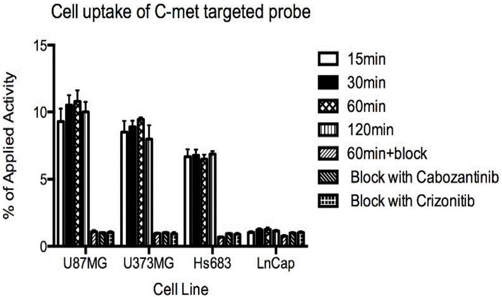
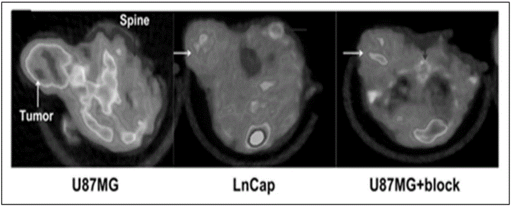
[0068] Establishment of nude mouse glioma (U87MG) subcutaneous xenograft tumor model, probe 68 One hour after Ga-NODAGA-cMET-1 injection, a 5-minute static PET imaging scan was performed, combined with a CT scan. The result is as Figure 4 . 68 Ga-NODAGA-cMET-1 was obviously aggregated in the tumor site 2 hours after intravenous injection, while the probe was not aggregated in the control group OVCAR3 tumor (negative expression of C-MET). After being blocked by a large amount of unlabeled NODAGA-cMET-1, the accumulation amount of the probe in...

Embodiment 3

[0072] 1) 18 Synthesis and characterization of F-cMET-2 probe;

[0073]

[0074] Reagent: 4 mg cMET-2, K 18 F, K 222 / K 2 CO 3 solution ( 18 For F labeling, containing 15mgK 222 and 3.5mgK 2 CO 3 per mL), dry DMSO.

[0075] Operation process:

[0076] use standard 18 F radioactive labeling reaction apparatus for labeling. Concretely, the K 18 F(K 2 CO 3 Neutralize H 18 F produced), add 1mLK 222 / K 2 CO 3 solution, add 1mL acetonitrile and stir, pass N 2 , heated to 85°C to dry the solvent, then repeatedly added 1 mL of acetonitrile and dried twice (3 times in total), to remove all K 18 water in F. Take 4mg of CMET-2 reactant, dissolve in 1mL of anhydrous DMSO, mix well and then N 2 Added into the reactor under protection, heated to 110°C for 30min. Purified by preparative HPLC 18 F-CMET-2. 1 H NMR (300MHz, Chloroform-d) δ8.78 (d, J=2.7Hz, 1H), 7.71(s, 1H), 7.24–7.21(m, 1H), 6.87(s, 1H), 5.35–5.19( m, 1H), 4.19(s, 2H), 3.83(d, J=28.6Hz, 4H), 3.30(d, ...


PUM


Abstract

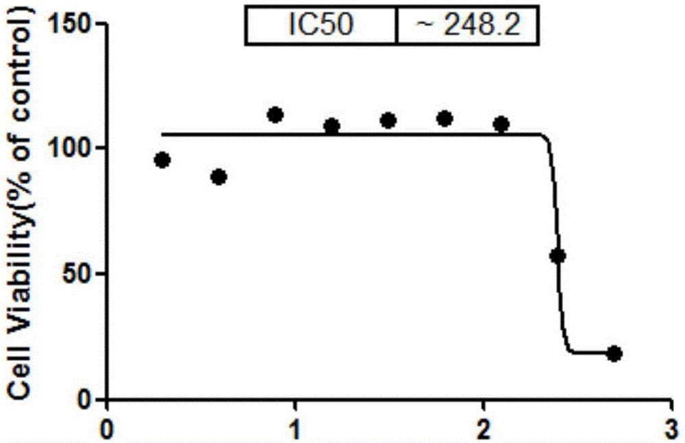
The invention especially relates to a radioactive C-MET-targeted affinity micromolecular compound and application thereof, belonging to the field of medical diagnosis and treatment. According to the invention, in-vivo tracking and imaging of diseases like tumors is realized through radiolabeling of a C-MET receptor antagonist. The radioactive C-MET-targeted affinity micromolecular compound can be used for monitoring the characteristics of biochemical changes of a hepatocyte growth factor receptor, visually displays the distribution and quantity of C-MET and is applicable to specific detection of a plurality of cancers including glioma, lung cancer, prostatic cancer, breast cancer, esophagus cancer, stomach cancer, liver cancer, pancreas cancer, ovarian cancer, rectal cancer and cervical cancer. The radioactive C-MET-targeted affinity micromolecular compound also has certain anticancer activity and has wide clinical application prospect.

Description

technical field [0001] The invention belongs to the field of medical diagnosis and treatment, in particular to a radioactive C-MET targeting affinity small molecular compound and its application. Background technique [0002] The encoded product of the proto-oncogene hepatocyte growth factor receptor (c-MET) is the cell membrane receptor of hepatocyte growth factor (HGF), which belongs to the tyrosine kinase receptor. The body dimerization leads to the activation of a series of signaling pathways, which lead to tumor angiogenesis, proliferation, enhanced cell motility and invasion, and finally metastasis. Therefore, the interaction between C-MET and its ligand HGF is closely related to the occurrence of various human tumors. , development related. Existing studies have found that C-MET receptors are highly expressed in a variety of tumors, including glioma, lung cancer, prostate cancer, breast cancer, esophageal cancer, gastric cancer, liver cancer, pancreatic cancer, ovari...

Claims


Application Information

Patent Timeline
IPC IPC(8): C07D215/38C07D401/12C07B59/00A61K51/04A61P35/00A61K101/02
CPCA61K51/0455C07B59/002C07B2200/05C07D215/38C07D401/12
Inventor 程震张翱申宝忠卜丽红陈浩
Owner 上海准视生物科技有限公司
Learn More