Method for calcification of cancer cells
A technology of cancer cells and seeding density, applied in the field of calcified cancer cells, can solve the problems of affecting mammalian cell activity and lack of cell wall protection, and achieve the effect of inhibiting cell activity and improving biological safety.
- Summary
- Abstract
- Description
- Claims
- Application Information
AI Technical Summary
Problems solved by technology
Method used
Image

Examples
Embodiment 1
[0034] 1. Preparation of targeting calcified cancer cells—A
[0035] Spread HeLa cells in a 24-well plate, the number of cells per well is about 2×10 5, when the cells grow to about 70% of the bottom area of the orifice plate, the calcification process is started. First pour off the culture medium of the cells, wash with PBS 1-2 times, then add DMEM medium containing 300 μg / mLFA to the well plate, place the cells in the incubator, and adjust the CO 2 The concentration is 5%, the air is 95%, the temperature is 37°C, after incubation for 15min, add 11.8mM Ca 2+ (including newly added 10mMCaCl 2 ) DMEM medium calcification solution, place the orifice plate in a cell culture incubator for calcification treatment for 30 minutes, absorb the calcification solution and add fresh calcification solution to continue the treatment for 60 minutes. The FA and calcification solution were treated alone as a control, and the solution was sucked off after the treatment to obtain calcified ...
Embodiment 2
[0047] Laser confocal microscopy, life-and-death staining and MTT assay were used to detect the killing effect of calcification treatment on cancer cells. The specific operation is as follows:
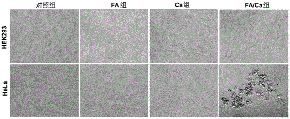
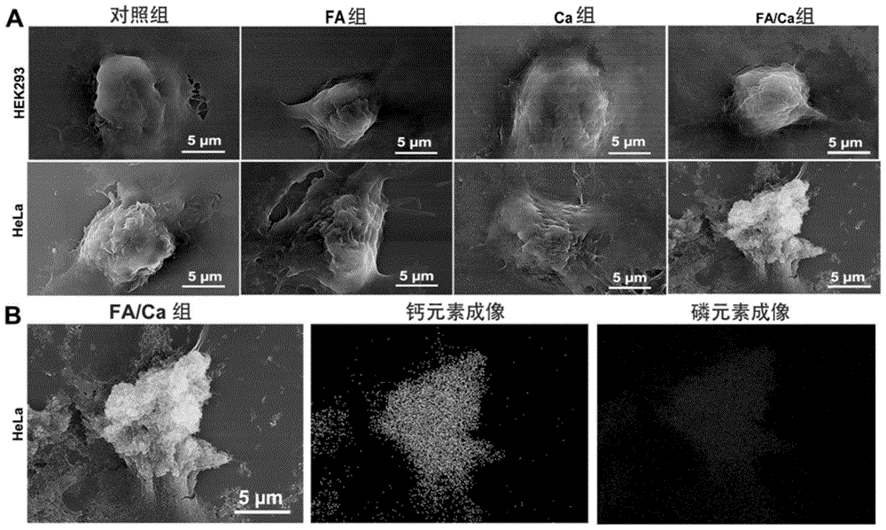
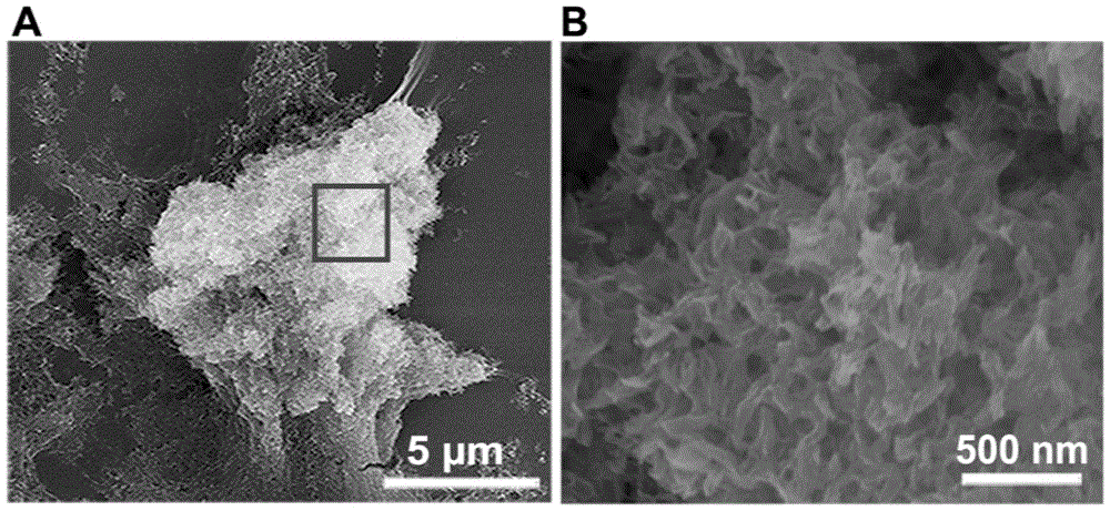
[0048] 1. Changes after cell calcification
[0049] Before calcification treatment, the cell membrane and nucleus of cancer cells were fluorescently stained with living cell dyes PKH26 and Hoechst33342, respectively. After the calcification treatment, the calcium-specific fluorescent dye calcein was used to fluorescently stain the mineral layer. After the staining was completed, the changes in cell morphology over time were observed under a laser confocal microscope. For the results, see Figure 5 .
[0051] (1) HEK293, MCF7 and HeLa cells were treated with 1×10 4 Cells / well were inoculated in 96-well plates respectively, and after 24 hours of culture, the cells were subjected to calcification treatment, and the calcification treatment method was the...
Embodiment 3
[0059] A HeLa tumor animal model was constructed to test the effect of targeted calcification in vivo. The operation and detection process is as follows:
[0060] (1) HeLa cell line with 5×10 6 The amount of one / only was inoculated into nude mice (20 nude mice, female) subcutaneously as a mouse drug resistance model, and after 10 days, the tumor volume grew to 80mm 3 , start the test.
[0061] (2) Inject 200 μL, 1 mg / mL folic acid intraperitoneally, wait for about 15 minutes, wait for the folic acid to accumulate in the tumor site, inject high calcium DMEM medium (50 mMCa 2+ ), injected once a day, treated continuously for 15 days, and observed the effect of tumor calcification. Injection of calcification solution and folic acid alone and blank group were used as controls.
[0062] (3) Use micro-CT technology to scan the tumor, and perform three-dimensional reconstruction after scanning to determine the result of tumor calcification; slice the calcified tumor, perform fluo...
PUM
 Abstract
Description
Claims
Application Information
 - Generate Ideas
- Intellectual Property
- Life Sciences
- Materials
- Tech Scout
- Unparalleled Data Quality
- Higher Quality Content
- 60% Fewer Hallucinations
Browse by: Latest US Patents, China's latest patents, Technical Efficacy Thesaurus, Application Domain, Technology Topic, Popular Technical Reports.
© 2025 PatSnap. All rights reserved.Legal|Privacy policy|Modern Slavery Act Transparency Statement|Sitemap|About US| Contact US: help@patsnap.com