Method for culturing umbilical cord mesenchymal stem cells in separated mode from umbilical cord outer layer amnion tissue
A technology of mesenchymal stem cells and outer amniotic membrane, which is applied in the field of isolation and culture of mesenchymal stem cells, can solve the problems of limited growth space of mesenchymal stem cells, influence on the separation, extraction, development and utilization of umbilical cord mesenchymal stem cells, and influence on the vitality of stem cells, etc.
- Summary
- Abstract
- Description
- Claims
- Application Information
AI Technical Summary
Problems solved by technology
Method used
Image
Examples
Embodiment 1
[0086] Example 1 Selection of serum-free medium for mesenchymal stem cells
[0087] (1) Content screening of serum substitutes
[0088] Test medium: 0.1 volume part of β-mercaptoethanol, 10ng / ml recombinant human basic fibroblast growth factor (b-FGF, Peprotech company), 1 volume part of non-essential amino acid aqueous solution (11140, Gibco company), 1, 2, 5, 8, 10, 12, 15 or 20 parts by volume of KnockoutFBS serum substitute (10828-028, Gibco), 89 parts by volume of a-MEM.
[0089] In the biological safety cabinet, take the third-generation hUC-MSC isolated from the umbilical cord Huatong glue tissue of the natural delivery neonates, and use them as 2×10 4 Cells / cm 2 The density is inoculated in T75 cell culture flask, and 12-15ml of conventional commercially available medium is added to culture cells. After culturing and observing that the cells have fully adhered to the wall, 15 mL of test medium was replaced. Observe the cell growth.
[0090] Results: In the three concentrati...
Embodiment 2
[0095] Example 2 Method for assisting extraction of umbilical cord mesenchymal stem cells with red blood cell lysate
[0096] Collection and transportation of samples: The umbilical cord samples of newborns delivered by cesarean section are collected under aseptic conditions and placed in the umbilical cord preservation and transport solution (D-Hank's solution) containing penicillin sodium, streptomycin sulfate, gentamicin and amphotericin B The final concentration of penicillin sodium, streptomycin sulfate and gentamicin in the protection solution is 150U / mL, and the final concentration of amphotericin B is 300U / ml), transported to the clean GMP cell laboratory on ice within 2 hours;
[0097] Cleaning and disinfection of samples: Put fresh umbilical cord samples in a 50ml sterile centrifuge tube in a biological safety cabinet, rinse with 75% alcohol twice, and rinse with sterile normal saline three times;
[0098] Pretreatment of umbilical cord tissue: Use ophthalmological scissor...
Embodiment 3
[0102] Example 3 Method for assisting extraction of umbilical cord mesenchymal stem cells with red blood cell lysate
[0103] The red blood cell lysate used contains 10g / L of NH 4 Cl and 0.1mMNa 2 -EDTA, pH is 7.2-7.4.
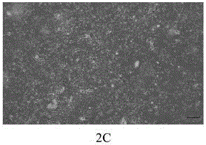
[0104] Refer to the method in Example 2. After culturing to the 5th day, the mesenchymal stem cells crawled out from the bottom of the tissue block. Around the 7th day, the mesenchymal stem cells formed a swirling cell cluster. After removing the tissue block and replacing the fresh medium, About 12 days, the cells reached 60% confluence and were passaged after trypsin digestion; after the third passage, the purity of the cells was greater than 99%, and the viability was greater than 95%.
PUM
 Abstract
Description
Claims
Application Information
 - R&D
- Intellectual Property
- Life Sciences
- Materials
- Tech Scout
- Unparalleled Data Quality
- Higher Quality Content
- 60% Fewer Hallucinations
Browse by: Latest US Patents, China's latest patents, Technical Efficacy Thesaurus, Application Domain, Technology Topic, Popular Technical Reports.
© 2025 PatSnap. All rights reserved.Legal|Privacy policy|Modern Slavery Act Transparency Statement|Sitemap|About US| Contact US: help@patsnap.com


