
System and method for diffused optical tomography and photoacoustic tomography combined measurement

A technology of photoacoustic tomography and optical tomography, used in echo tomography, diagnostic recording/measurement, medical science, etc. Effects of quality optical parametric imaging capabilities

Inactive Publication Date: 2015-09-02
TIANJIN UNIV
View PDF2 Cites 13 Cited by
  • Summary
  • Abstract
  • Description
  • Claims
  • Application Information

AI Technical Summary

Problems solved by technology

However, due to the high scattering characteristics of tissue, this type of pure optical imaging technology cannot have both imaging depth and spatial resolution, and its spatial resolution is limited to 5%-10% of the imaging tissue size; Photoacoustic tomography (PAT) method effectively combines the advantages of optical imaging and ultrasound imaging, and is a biophoton imaging method using ultrasound as a medium.

Method used


Image

  • System and method for diffused optical tomography and photoacoustic tomography combined measurement
  • System and method for diffused optical tomography and photoacoustic tomography combined measurement
  • System and method for diffused optical tomography and photoacoustic tomography combined measurement

Examples


Embodiment 1

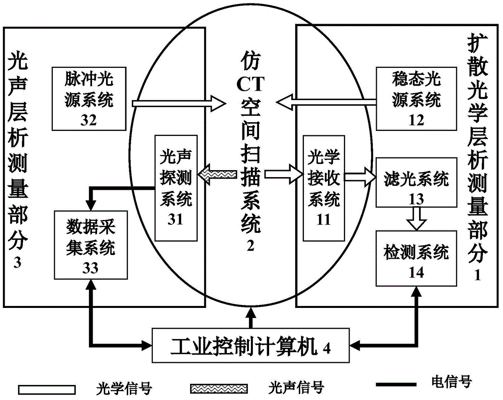
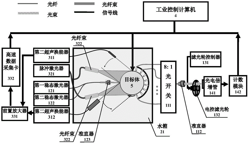
[0028] Such as figure 2 As shown, the specific implementation case 1 of the present invention is a joint measurement system structure composed of a dual-wavelength dual-bandwidth diffusion optical tomography measurement part and a photoacoustic tomography joint measurement part.

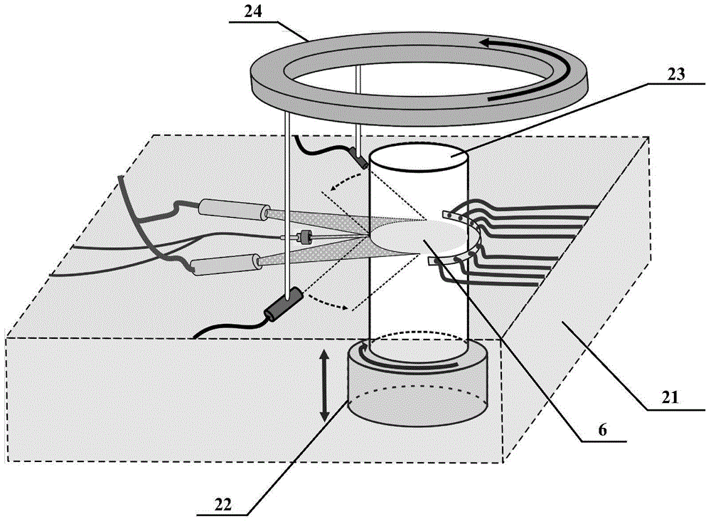
[0029] Diffuse optical tomography measurement part implementation process (such as figure 1 with figure 2 shown): two first stable semiconductor lasers (121) and second stable semiconductor lasers (122) (average powerfigure 1 with figure 2 As shown): the pulsed light source system adopts a distributed multi-beam incident mode to achieve uniform irradiation of high-energy incident light on the surface of the target (5). The pulsed light source system (32) adopts a pulsed laser (321) (OPO laser pumped by a Q-switched Nd:YAG laser (wavelength 532nm), with a wavelength tunable range of 600-900nm), and it is planned to select two wavelengths close to the DOT measurement part , pulse width ≤ 10ns, re...

Embodiment 2

[0032] The combined measurement of diffusion optical tomography and photoacoustic tomography of the biological tissue model was performed using the system described in Example 1. The system can realize high-contrast and high-spatial-resolution imaging of the lesion area, monitor tumor neovascularization and blood oxygenation, and observe endogenous or exogenous specific markers in vivo. For example, when indocyanine green (ICG) is used as an exogenous absorption contrast agent in small living animal experiments, the working wavelength λ 1 The peak wavelength of the absorption spectrum according to ICG was set to 785 nm. In addition, in order to effectively obtain the blood oxygen information in the tissue, the system working wavelength λ 2 According to the extinction point of hemoglobin (about 805nm), it is planned to be set to 830nm. During measurement, the target to be measured is placed in a cylindrical imaging cavity, and the gap between the imaging cavity and the target...


PUM

PropertyMeasurementUnit
Diameteraaaaaaaaaa

Abstract

The invention discloses a system and method for diffused optical tomography and photoacoustic tomography combined measurement. The system comprises a diffused optical tomography measurement part (1), an imitating CT space scanning system (2), a photoacoustic tomography measurement part (3) and an industrial control computer (4), wherein the diffused optical tomography measurement part (1) and the photoacoustic tomography measurement part (3) are jointly connected with the imitating CT space scanning system (2) to form an integrated multi-mode measurement system, and system automatic control software of the industrial control computer (4) is used for achieving automatic detection and data collection of a diffused optical signal and a photoacoustic signal. Compared with the prior art, by means of the system and method for the diffused optical tomography and photoacoustic tomography combined measurement, the defects of an existing single-mode imaging technique are overcome, the defect that for the prior art, the spatial resolution and the quantitative feature cannot be achieved simultaneously is overcome, the imaging method can achieve the spatial resolution of the photoacoustic tomography technique and the quantitative precision of the diffused optical tomography technique, and therefore the high-quality optical parameter imaging function is achieved.

Description

technical field [0001] The invention relates to the field of application of optical imaging technology in the direction of biomedicine, in particular to a combined measurement system of diffusion optical tomography and photoacoustic tomography and a multimodal measurement method. Background technique [0002] With the development of biology, medicine, optoelectronic technology and computer technology, various advanced medical imaging devices are constantly emerging, providing medical images of various modes for clinical diagnosis. Due to the different imaging mechanisms and the high complexity of human tissue structure, medical images of a single mode cannot provide enough information for doctors, and the information provided by medical images of different modalities cannot cover and complement each other. Therefore, in clinical diagnosis and treatment, it is of great significance to the development of multimodal medical imaging technology. [0003] With the development of ...

Claims


Application Information

Patent Timeline
IPC IPC(8): A61B5/00A61B6/03
CPCA61B5/0095A61B5/0033A61B5/0073A61B5/72A61B6/035
Inventor 李娇王艺涵高峰解陶然张燕
Owner TIANJIN UNIV
Learn More