A detection method and application of degraded dna from spoilage material

A detection method and technology for testing materials, applied in biochemical equipment and methods, microbial determination/inspection, etc., can solve problems such as allele loss and low detection rate of loci, and achieve accurate screening results and high forensic application value. Effect

Active Publication Date: 2017-05-24
HEBEI MEDICAL UNIVERSITY
View PDF1 Cites 0 Cited by
  • Summary
  • Abstract
  • Description
  • Claims
  • Application Information

AI Technical Summary

Problems solved by technology

[0003] The purpose of the present invention is to provide a detection method and application thereof for the degraded DNA of corrupted specimens, to provide a new detection technology for degraded DNA of corrupted specimens, to solve the problem of existing detection methods for detecting degraded DNA of corrupted specimens. Problems with missing alleles and low detection rates of loci

Method used


Image

  • A detection method and application of degraded dna from spoilage material
  • A detection method and application of degraded dna from spoilage material
  • A detection method and application of degraded dna from spoilage material

Examples


Embodiment 1

[0025] Example 1 Acquisition of DNA genetic markers located in the nucleosome core region

[0026] Its DNA genetic markers include SNVs (single nucleotide variation sites), Indels (insertion-deletion polymorphism sites), and STRs (short tandem repeat polymorphism sites).

[0027] experimental method:

[0028] (1) Collect white blood cells from the peripheral blood of healthy people, use micrococcal nuclease (micrococcal nuclease, MNase) to digest the white blood cells after blood lysis; add CaCl at a final concentration of 1mM 2 The solution and micrococcal nuclease with a final concentration of 3U / ul were incubated at 37°C for 3 hours to release the nucleosome core, and the DNA in the digested white blood cells was extracted using the phenol-chloroform method to obtain the nucleosome core in human blood tissue Region DNA;

[0029] (2) Use the Illumina Hiseq 2000 Genome Analyzer analysis platform to sequence the obtained nucleosome core region DNA;

[0030] (3) Use bwa (0.6...

Embodiment 2

[0040] Example 2 Designing primers by taking partial labeling of the obtained nucleosome core region as an example

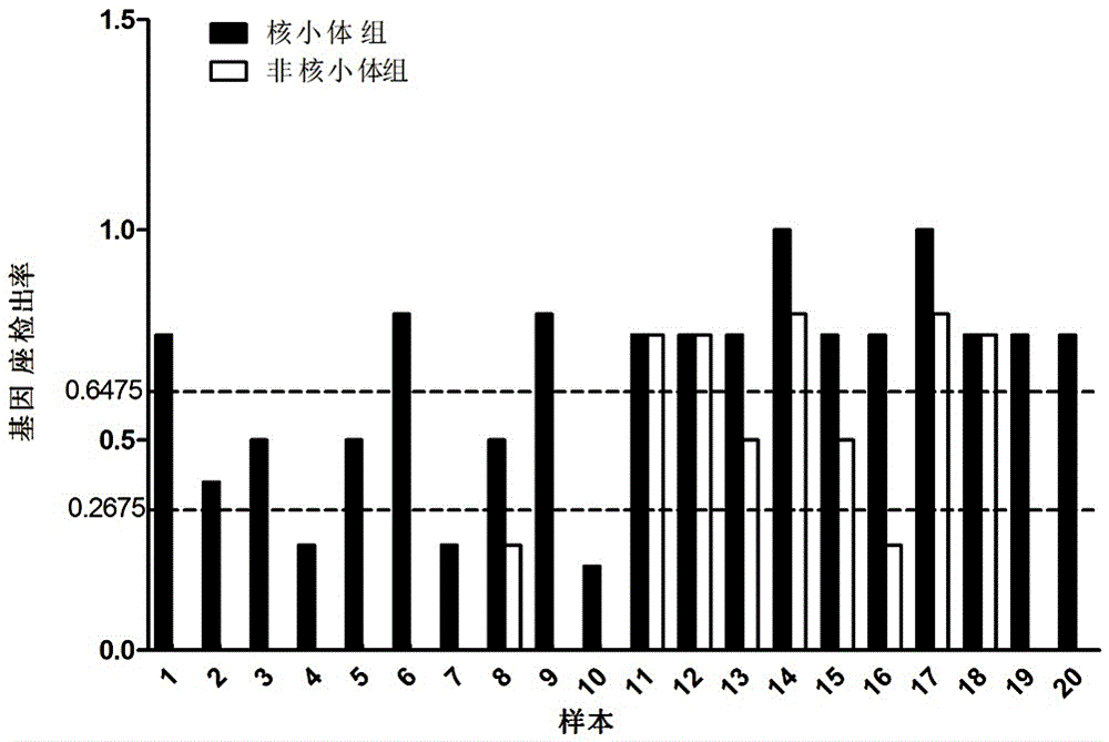
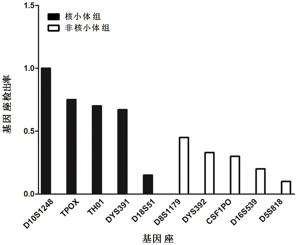
[0041] The STRs marker data obtained in Example 1 were compared with 44 commonly used genetic markers in forensic science. There were 5 STR loci successfully matched, and these 5 STR loci were selected as nucleosome group STRs, namely D10S1248, D18S51, TH01, TPOX and DYS391, see the markers marked with two asterisks in Table 3. Five STR loci were randomly selected from the remaining 39 loci on the unalignment as non-nucleosome group STRs, namely CSF1PO, D5S818, D8S1179, D16S539 and DYS392, see the marker marked with an asterisk in Table 3. Details of selected genetic markers commonly used in forensic science located in different regions of nucleosomes are given in Table 3. By redesigning the primers, the lengths of the amplified fragments of the above 10 selected STRs are all less than the length of the nucleosome core fragment of 147bp. The information of the ...

Embodiment 3

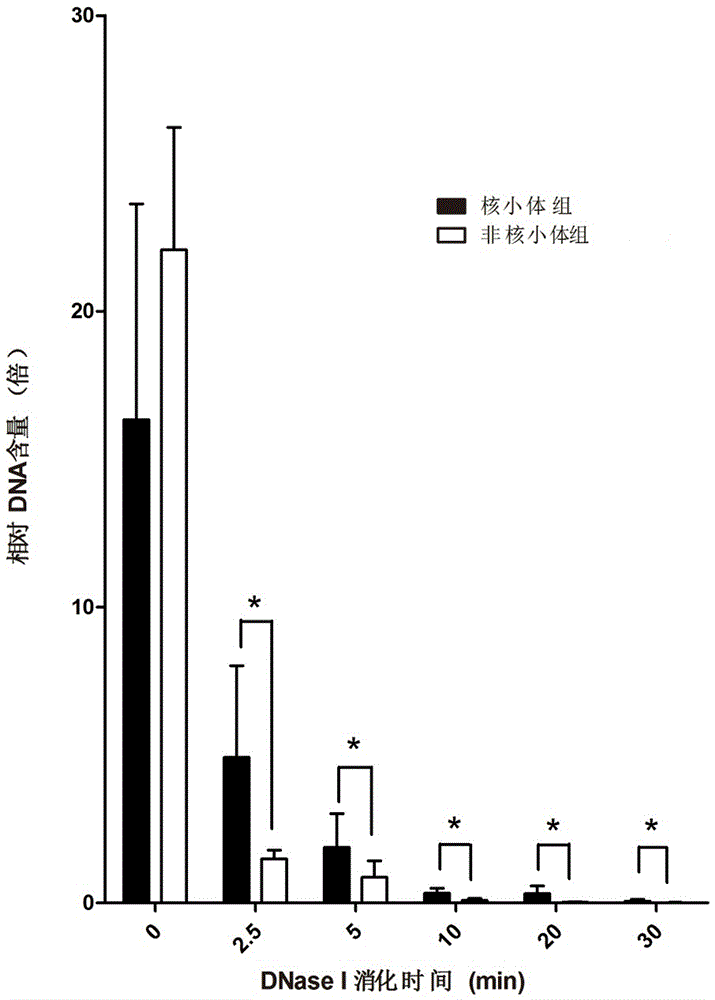
[0048] Example 3 Manual preparation of degraded samples and extraction of degraded DNA

[0049] The blood samples of 5 different individuals (A, B, C, D and E) were taken as samples, and the DNA in the blood was extracted respectively (using QIAamp DNA Blood Midi kit, Qiagen, Germany), and 10 µg of DNA was taken at a concentration of 0.01 U / μl of the digestive enzyme DNase I for digestion, and after digestion to 0, 2.5 min, 5 min, 10 min, 20 min and 30 min each time point, samples were taken, and 30 samples of artificially degraded samples were obtained.


PUM

PropertyMeasurementUnit
correlation coefficientaaaaaaaaaa

Abstract

The invention discloses a detection method for degraded DNA of a corrupt sample material, comprising the following steps: a) obtaining DNA located in the core region of nucleosome of human blood tissue; b) sequencing the DNA in the core region of the nucleosome; The sequence obtained by sequencing is compared with the human genome sequence information, and screening is carried out to obtain the SNVs, Indels or STRs genetic markers in the core region of the nucleosome; d) Design upstream and downstream primers according to the sequences of the SNVs, Indels or STRs genetic markers; e) Extract the DNA of the spoiled specimen; f) Use the upstream and downstream primers to amplify the DNA of the spoiled specimen by PCR to obtain an amplification product; g) Electrophoresis the amplified product, and obtain the amplification product A genotyping analysis was performed for each locus. The invention uses the genetic markers in the core region of the nucleosome to detect the degraded DNA of the corrupted sample, the detection rate of the locus is higher, and the obtained detection information is more comprehensive, so that it can be more accurate in forensic identification and archaeological analysis. for accurate and reliable conclusions.

Description

technical field [0001] The invention relates to the field of forensic DNA detection, in particular to a method for detecting degraded DNA of corrupt specimens and its application. Background technique [0002] In the detection of death and injury cases, corrupt biological samples are often encountered. Because the DNA in the samples is severely degraded, if the conventional kit is used for detection, the results obtained will be incomplete or poor in repeatability. The spectrum can be seen with ladder bands and Stutter bands, unbalanced allelic amplification, loss of loci, weak signal of amplified loci, and no way to determine the type, etc., resulting in a low detection rate of such cases. At present, although MiniSTR and SNP amplification systems commonly used to detect degraded DNA can solve some problems, there are still allele loss, low detection rate of loci, and poor comprehensiveness and accuracy of detection information obtained. The problem is also the technical b...

Claims


Application Information

Patent Timeline
Patent Type & Authority Patents(China)
IPC IPC(8): C12Q1/68
Inventor 丛斌董春楠李淑瑾马春玲
Owner HEBEI MEDICAL UNIVERSITY
Why Eureka
Try Eureka