
Procalcitonin detection kit and detection method

A detection kit, procalcitonin technology, applied in biological tests, measuring devices, material testing products, etc., can solve the problems of unstable detection results, poor quantitative accuracy, long time required, etc., and achieve clear clinical guiding significance , fast response, easy to operate

Active Publication Date: 2017-03-15
WEIHAI NEOPROBIO
View PDF5 Cites 1 Cited by
  • Summary
  • Abstract
  • Description
  • Claims
  • Application Information

AI Technical Summary

Problems solved by technology

Among them, high-performance liquid chromatography is time-consuming and difficult to automate; ELISA method has poor quantitative accuracy, long operation time, and low degree of automation, and is mostly used for qualitative detection; radioimmunoassay has a sensitivity of 4pg / ml, which can detect normal human serum PCT. The double-antibody sandwich method is sensitive, but the disadvantage is that it takes a long time, the test results are unstable, the repeatability is worse than ELISA, and there is a risk of radioactive contamination; latex particle-enhanced immune turbidimetry (PETIA) can be easily applied and fully automatic biochemical instrument, but the sensitivity is low, and the cut-off value of 0.5ng / ml for procalcitonin diagnosis of systemic infection (sepsis) cannot be accurately detected; the sensitivity of the gold standard method is low, generally only qualitative, not quantitative, especially The shortcoming of poor repeatability limits its clinical application, especially not suitable for the quantitative detection of body fluid marker proteins that need accurate quantification to help diagnose diseases; immunoluminescence method has strong specificity and high sensitivity, but requires Expensive equipment and experienced operators are generally used in specific medical institutions

Method used

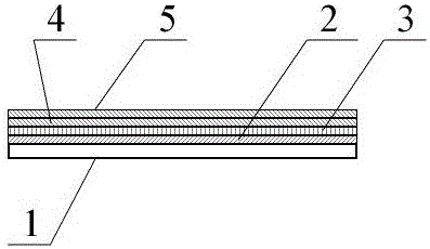

Image

  • Procalcitonin detection kit and detection method
  • Procalcitonin detection kit and detection method
  • Procalcitonin detection kit and detection method

Examples


Embodiment 1

[0033] The various components of the test paper card in the procalcitonin detection kit can be prepared by the following measures:

[0034] 1. Preparation of sample pad 2:

[0035] Soak the glass fiber membrane in the treatment solution containing 2.0% TritonX-100, 3% BSA, 0.2MTris buffer, pH7.5, soak at 4°C for 4 hours, then place it in an oven, and dry it at 37°C for 2 hours .

[0036] 2. Preparation of binding pad 3 for absorbing fluorescent microsphere-labeled antibody:

[0037] Soak the glass fiber membrane in 200mM Tris-HCL treatment solution (containing 2.0% Triton X-100, 3.0% BSA, pH7.5), soak at 4°C for 4 hours, then take it out of the oven at 37°C and dry it for 4 hours, and set it aside. Put the glass fiber membrane on the Bio-DotXYZ3050 three-dimensional spraying platform, use the Bio-Jet Quanti300 non-contact micro-quantitative nozzle to spray the rare earth fluorescent microsphere-labeled procalcitonin monoclonal antibody on the glass fiber membrane, and dry it...

Embodiment 2

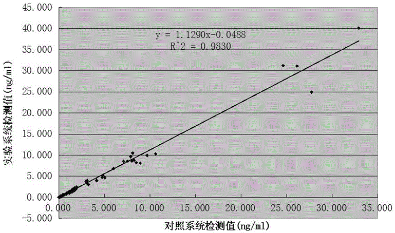
[0046] Embodiment 2: accuracy test

[0047] Select the above test paper card and fluorescence immunochromatography analyzer (model: NEO-007),

[0048] Setting of the parameters of the fluorescence immunoassay analyzer: after setting the process parameters of the test paper card on the fluorescence immunoassay The original calcitonin calibrator is measured with a test paper card to obtain the fluorescence intensity value of each calibrator, and the result is input into the parameters of the analyzer to complete the setting of the parameters of the analyzer.

[0049] Main testing materials: clinical samples were obtained from relevant hospitals, a total of 200 electrochemiluminescence immunoassay value samples, including 100 serum samples, 100 whole blood samples, and the distribution range of procalcitonin content was between 0-40ng / mL.

[0050] Detection method:

[0051] Step 1: Equilibrate the detection reagent and sample to room temperature, take out the test paper card, a...

Embodiment 3

[0058] Embodiment 3: precision test

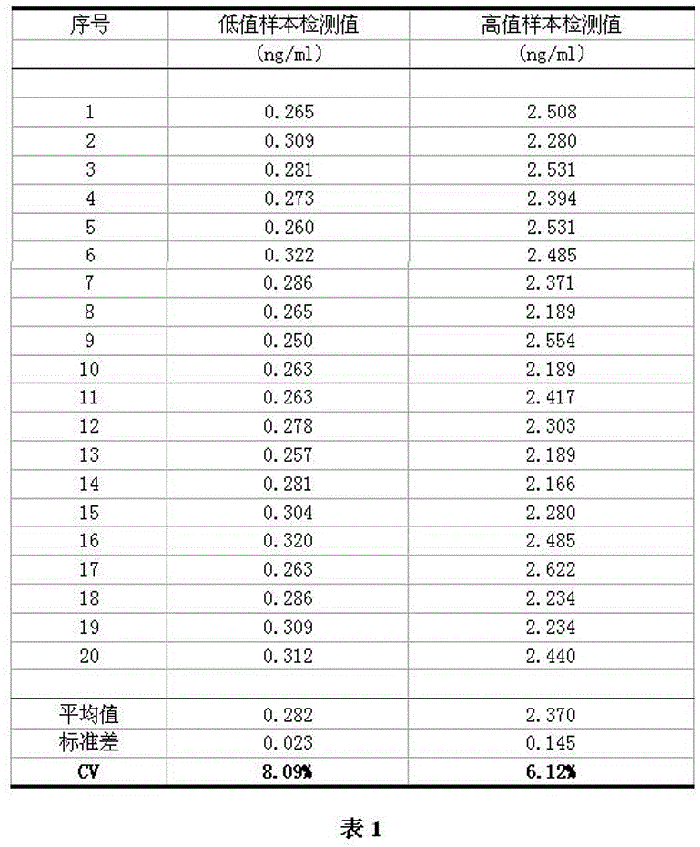
[0059] Using the test paper card and measuring system of Example 2, the test paper card and the fluorescent immunochromatographic analyzer of the present invention were tested for precision.

[0060] Main testing materials: clinical samples obtained from relevant hospitals, a total of 2 serum samples with chemiluminescence immunoassay value, among which the clinical measurement value of the low value fixed value sample is 0.26ng / ml, and the clinical measurement value of the high value fixed value sample is 2.28ng / ml .

[0061] Detection method:

[0062] Using the test paper card and measuring system of Example 2, each of the 2 fixed-value samples was repeatedly measured 20 times.

[0063] Analysis of test results:

[0064] After the clinical sample test reagents are prepared, the clinical samples are tested according to the test method, and the test results are analyzed.

[0065] test results:

[0066] as attached image 3 As shown i...


PUM

PropertyMeasurementUnit
diameteraaaaaaaaaa
diameteraaaaaaaaaa

Abstract

The invention relates to the field of fluorescence immunochromatography technique in medical immunology, specifically to a procalcitonin detection kit and a procalcitonin detection method. The detection kit is provided with a test cassette and is characterized in that the test cassette is successively provided with, from bottom to top, a PVC plate, a sample pad, a combination pad, a cellulose nitrate film and a water-absorbing pad, wherein a procalcitonin monoclonal antibody labeled by a rare earth fluorescent microsphere is adsorbed on the combination pad, the rare earth fluorescent microsphere has a diameter of 60 to 120 nm, is doped by rare earth lanthanide, is stable in a ground state and emits fluorescent light with a wavelength in a range of 540 to 600 nm under the action of an excitation light source in a wavelength range of 340 to 380 nm, and the monoclonal antibody is a purified mixed monoclonal antibody and is originated from monoclonal antibody cell strains directed at 2 to 6 different procalcitonin antigen epitopes. The procalcitonin detection kit has the advantages of simple operation, rapid reaction, high sensitivity, strong specificity, etc.

Description

technical field [0001] The invention relates to the technical field of fluorescent immunochromatography in medical immunology, in particular to a procalcitonin detection kit and a detection method capable of rapidly and accurately quantitatively analyzing procalcitonin. Background technique [0002] Procalcitonin (PCT) is a glycoprotein composed of 116 amino acids with a molecular weight of 13KD. It is the precursor peptide of calcitonin (CT) and can exist in normal human serum in free form. In 1993, Lancet reported for the first time that the level of procalcitonin was significantly increased in sepsis. When the blood PCT concentration was greater than 0.5ng / ml, it indicated the possibility of sepsis; if it was greater than 2ng / ml, it indicated the existence of sepsis; Sepsis or shock, so PCT can be used as a specific indicator for the diagnosis of sepsis. In recent years, with the deepening of research, it has been found that in the early stage of bacterial infection, the...

Claims


Application Information

Patent Timeline
Patent Type & Authority Patents(China)
IPC IPC(8): G01N33/74G01N33/577G01N33/544C07K16/26
CPCC07K16/26G01N33/544G01N33/74
Inventor 李红江孙宁宁刘衍亮
Owner WEIHAI NEOPROBIO
Learn More