In-vitro culture method of visual cortex neuron

A technique for in vitro culture of neurons

Inactive Publication Date: 2015-05-13
THE FIRST AFFILIATED HOSPITAL OF THIRD MILITARY MEDICAL UNIVERSITY OF PLA
View PDF4 Cites 3 Cited by
  • Summary
  • Abstract
  • Description
  • Claims
  • Application Information

AI Technical Summary

Problems solved by technology

However, such a culture system has not been reported in China, and similar foreign research systems are mostly established on the hippocampus, and there has been no report on the establishment of an in vitro neuron culture system for the study of synaptic plasticity of visual cortex neurons.

Method used


Image

  • In-vitro culture method of visual cortex neuron
  • In-vitro culture method of visual cortex neuron
  • In-vitro culture method of visual cortex neuron

Examples


Embodiment 1

[0044] Embodiment 1 Utilizes the culture method described in the present invention to cultivate neurons

[0045] 1. Culture plate treatment:

[0046] Place a circular cover glass with a diameter of 14mm in advance in a 24-well plate, add 0.05g / L polylysine for coating, overnight, absorb the polylysine, rinse with sterile ultrapure water for 3 times, and dry at room temperature Dry, add 2mg / L laminin to coat for 2h.

[0047] 2. Neuron culture:

[0048] (1) Material collection: female rats were killed by neck dislocation, and the abdomen was disinfected with 75% alcohol to obtain fetuses by laparotomy. Take out the fetal mouse and take its head. Ophthalmic tweezers were used to fix the head of the mouse, and the corneal scissors were cut from the foramen magnum along both sides of the skull to the eyeball. The skull was removed, and the brain was removed. The visual cortex is located in the dorsal area of ​​the occipital area of ​​the brain, with a size of about 2mm*3mm and ...

Embodiment 2

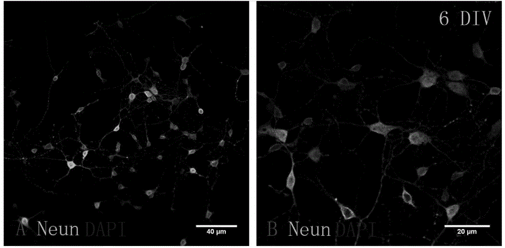
[0050] Embodiment 2 uses rat visual cortex neuron cell line of the present invention to carry out research on synaptic plasticity of visual cortex neurons

[0051] 1. Identification of the purity of visual cortex neurons

[0052] method:

[0053] Take the rat visual cortex neuron slides that were cultured for 6 days according to the steps (1) and (2) of Example 1, rinse 2 times with 0.01M PBS, 3min / time; treat with 4% paraformaldehyde for 15min, rinse 4 times with 0.01M PBS , 5min / time; 0.25% triton-100 breakthrough treatment for 15min, 0.01M PBS rinse 4 times, 5min / time; add blocking solution containing 1% BSA and 10% rabbit serum, block at 37°C for 1h; Mouse Neun primary antibody (1:500), placed in a humid box, incubated overnight at 4°C. (5) Rinse 3 times with 0.01M PBS, 5min each time; add rabbit anti-mouse Alexa Fluor 647-marked secondary antibody (1:800), place in a wet box, incubate at 37°C for 45mins in the dark, rinse with 0.01M PBS in the dark 2 times, 5min / time; ...


PUM

PropertyMeasurementUnit
Diameteraaaaaaaaaa
Resistanceaaaaaaaaaa

Abstract

The invention relates to a neuron in-vitro culture method for synaptic plasticity research on a visual cortex neuron. The method is characterized by comprising the following steps: (1) carrying out pretreatment on cerebral tissues of fetal rats, separating visual cortex tissues and carrying out proteinase digestion and centrifugation; (2) adding an inoculation medium to terminate digestion; (3) blowing a single-cell suspension, sieving and counting; and (4) inoculating with the inoculation medium of which the density is 2.5*10<4> / cm<2> to 5*10<4> / cm<2>, carrying out adherent culture in a full liquid-exchange manner, and after cell attachment, carrying out subsequent culture in a half liquid-exchange manner. The invention further provides a visual cortex neuron cell line of rats, which is cultured by the neuron in-vitro culture method, and an application of the visual cortex neuron cell line in synaptic plasticity research on the visual cortex neuron. The culture method is efficient and simple; the neuron is high in purity, and good in state, and has a structural basis of neuronal synaptic plasticity; the functional state of the neuronal synaptic plasticity can be reflected; and the culture method can be applied to research on the synaptic plasticity of visual cortex of rats.

Description

technical field [0001] The invention relates to the field of cell engineering, in particular to a method for culturing visual cortex neurons in vitro. Background technique [0002] The primary visual cortex (brodmman 17 area), located in the posterior part of the cerebral hemisphere near the cerebellum, is an important part of the higher visual center. After birth, the visual nervous system of humans and mammals can change its original neural connections and synaptic structures according to the visual environment. The most sensitive period for this change is called the critical period of visual cortex plasticity. In humans, this period is postnatal 2 -10 years old, rats are 2-6 weeks after birth. Abnormal visual experience during the period will cause vision loss, but there are no organic lesions in the eye, which is a common amblyopia in ophthalmology. The lesion is mainly in the visual cortex, which is closely related to the plasticity of the visual cortex. During the c...

Claims


Application Information

Patent Timeline
IPC IPC(8): C12N5/0793C12Q1/02G01N33/569C12R1/91
Inventor 郑莎黎一鸣赵从建王浩姚军平袁侨英余涛阴正勤
Owner THE FIRST AFFILIATED HOSPITAL OF THIRD MILITARY MEDICAL UNIVERSITY OF PLA
Why Eureka
Try Eureka