Nucleic acid nano structure carrier-precious metal photosensitive contrast agent composite for living organism photo-acoustic imaging, preparation method and applications thereof
A nucleic acid nanometer and precious metal technology, which is applied to preparations for in vivo tests, medical preparations without active ingredients, and medical preparations containing active ingredients, etc., to achieve the effect of high-sensitivity photoacoustic imaging capabilities
- Summary
- Abstract
- Description
- Claims
- Application Information
AI Technical Summary
Problems solved by technology
Method used
Image
Examples
Embodiment 1
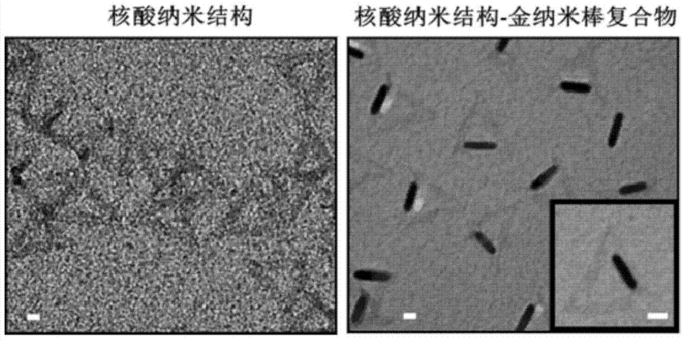
[0062] Example 1 Preparation of nucleic acid nanostructure carrier, noble metal photosensitive contrast agent and nucleic acid nanostructure carrier-noble metal photosensitive contrast agent complex
[0063] Mix 50 μL of M13 phage genomic DNA with a final concentration of 5 nM and 100 μL of staple strand and capture strand with a final concentration of 50 nM in 1 mL of 1×TAE / Mg 2+ (pH=8.0) solution, starting from 95° C., annealing at 20° C. gradually, annealing incubation time is 12-24 hours, and preparing a nucleic acid nanostructure with a pre-designed geometric shape. The triangular DNA origami used in this experiment was modified based on Rothemund's 2006 design (Nature, 2006, 440, 297-302.), and the capture strand was extended at 7 positions (5'-AAAAAAAAAAAAAAAA-original staple strand sequence -3'), its capture sequence is complementary to the sequence used by subsequent DNA modification gold rods.
[0064] Regulation of AgNO by seed-induced growth method using cetyltrim...
Embodiment 2
[0075] Example 2 Living Cell Uptake of Nucleic Acid Nanostructure-Gold Nanorod Complex
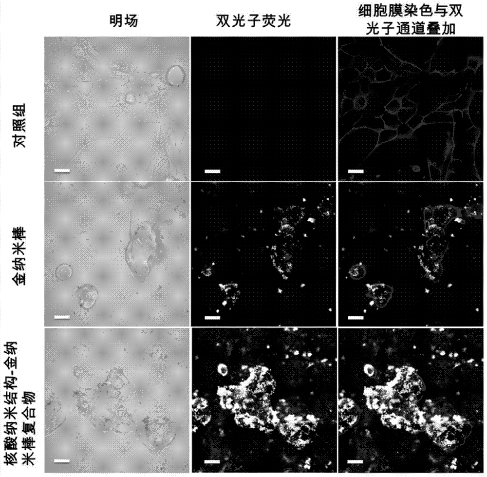
[0076] Mouse breast cancer 4T1 cells were seeded at 100mm 2 In a petri dish, use RPMI1640 medium (10% fetal bovine serum and double antibody) in 5% CO 2 Incubator, cultivate at 37°C. When the cells grow to about 80% confluence, digest the cells and inoculate in 35mm 2 The confocal small dishes were cultured overnight, and gold rods, DNA origami-gold nanorod composite structures (0.1 nM, diluted in complete medium) and the control group were added and incubated for 24 hours. The drug solution of each group was discarded, the cells were washed with PBS, and then observed with a two-photon fluorescence microscope with an excitation wavelength of 800 nm.
[0077] Observation results such as image 3 As shown, the pictures from top to bottom are the control group, the gold nanorod treatment group and the DNA origami-gold nanorod composite structure treatment group respectively, and from lef...
Embodiment 3
[0078] Example 3 In vivo photoacoustic imaging of nucleic acid nanostructure-gold nanorod complexes
[0079] Culture 4T1 cells to the logarithmic growth phase, digest with trypsin, collect the cells, adjust the concentration of the cell suspension to 1×10 7100 μL was orthotopically inoculated subcutaneously on the back of 5-6 week old female BALB / c nude mice to model subcutaneous mammary gland xenografts. After 1 week of modeling, when the tumor size reaches 100mm 3 , divided into two groups for administration treatment, namely the gold nanorod group (3nM×150 μL), the nucleic acid nanostructure-gold nanorod complex group (3nM×150 μL), and the drugs were injected intravenously. At 0, 3, and 24 hours after injection, in vivo photoacoustic imaging was performed on tumor-bearing mice using a small animal photoacoustic imaging system.
[0080] Figure 4 Shown are the photoacoustic imaging results of tumor-bearing mice at 0h, 3h and 24h after injection of gold rods. Depend on ...
PUM
 Abstract
Description
Claims
Application Information
 - R&D
- Intellectual Property
- Life Sciences
- Materials
- Tech Scout
- Unparalleled Data Quality
- Higher Quality Content
- 60% Fewer Hallucinations
Browse by: Latest US Patents, China's latest patents, Technical Efficacy Thesaurus, Application Domain, Technology Topic, Popular Technical Reports.
© 2025 PatSnap. All rights reserved.Legal|Privacy policy|Modern Slavery Act Transparency Statement|Sitemap|About US| Contact US: help@patsnap.com


