Immune chromatography fluorescence reagent strip for detecting cardiac troponin and preparation method thereof
A technique of cardiac troponin and immunochromatography, which is applied in the field of clinical medical testing to achieve the effects of saving costs, saving materials, and shortening detection time
- Summary
- Abstract
- Description
- Claims
- Application Information
AI Technical Summary
Problems solved by technology
Method used
Image
Examples
Embodiment 1
[0024] Embodiment 1 Preparation of tracer particle binding pad
[0025] Spray the tracer particle-labeled antibody / antigen marker evenly on the processed sample pad, and dry it at 25°C for 4 hours. The preparation of the tracer particle can be divided into the preparation of antibody IgG-fluorescein and the preparation of metal particle-antibody - Two-step preparation of the fluorescent complex:
[0026] A. Preparation of antibody IgG-fluorescein: Dissolve antibody IgG in 20-100Mmol PBS buffer, dissolve activated fluorescein (FITC, Cy5, etc.) in dimethyl sulfoxide (DMF), then mix antibody IgG with fluorescein Mix at 1:1~1:3, fully stir and react for 1 hour, then place the solution in a dialysis bag for gradient fluid dialysis for 3 times, each time for 6~8 hours, and the obtained solution is the antibody IgG-fluorescein complex;
[0027] B. Preparation of metal particle-antibody-fluorescent complex: Mix 50~200nm nanometer metal particles and antibody IgG-fluorescein at a ra...
Embodiment 2
[0028] Embodiment 2 Preparation of an immunochromatographic fluorescent reagent strip for detecting cardiac troponin
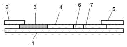
[0029] An immunochromatographic fluorescent reagent strip for detecting cardiac troponin, comprising a bottom plate 1, on which a sample pad 2, a tracer particle binding pad 3, a nitrocellulose membrane 4 and a water-absorbing pad 5 are sequentially pasted; Points 2-5 need to overlap by 1~2mm to ensure that the test sample passes through the test area and reaches the absorbent pad smoothly. The preparation method of each component is as follows:
[0030] 1) Preparation of sample pad: The sample pad can be made of glass fiber or polyester, and 5mg / ml of protein (BSA, casein, etc.) can be dissolved in buffer (PBS, Tris or glycine, etc.), and then add a small amount of surface activity agent (Tween20, etc.), adjust the pH to 6-8, soak the glass fiber or polyester material for 2-4 hours, take it out and dry it at 25°C for 8 hours;
[0031] 2) Preparation of trace...
Embodiment 3
[0037] Example 3 Detection of cardiac troponin protein using the reagent described in this utility
[0038] In embodiment 2 such as figure 1 The shown reagent strips are cut into 5mm widths and installed on a suitable reagent strip support shell, and the sample pad 2 faces the sample injection hole, and presses the shell tightly. Then the sample detection operation steps are as follows: (1) collect serum, plasma or whole blood samples and return to room temperature; (2) drop 100ul of the sample to be tested onto the sample pad 2 for reaction; (3) read the results, and after 90s The above reagent strips are placed in the immunoquantitative analyzer to read the results of the corresponding samples.
PUM
| Property | Measurement | Unit |
|---|---|---|
| Particle size | aaaaa | aaaaa |
| Thickness | aaaaa | aaaaa |
Abstract
Description
Claims
Application Information
 - R&D Engineer
- R&D Manager
- IP Professional
- Industry Leading Data Capabilities
- Powerful AI technology
- Patent DNA Extraction
Browse by: Latest US Patents, China's latest patents, Technical Efficacy Thesaurus, Application Domain, Technology Topic, Popular Technical Reports.
© 2024 PatSnap. All rights reserved.Legal|Privacy policy|Modern Slavery Act Transparency Statement|Sitemap|About US| Contact US: help@patsnap.com


