
Laser scanning fluorescence confocal microscopic endoscopic imaging system

A confocal microscopy imaging and laser light source technology, which is applied in the field of fluorescence confocal microscopy imaging systems, can solve the problems that fluorescence confocal microscopy imaging systems do not have the ability to quickly and accurately find targets, the time for three-dimensional tomographic images is long, and it is difficult to accurately image target objects. Achieve the effect of being beneficial to industrial application, reducing system cost and solving long scanning time

Active Publication Date: 2014-07-16
JIANGSU TIANNING PHOTON TECH
View PDF6 Cites 19 Cited by
  • Summary
  • Abstract
  • Description
  • Claims
  • Application Information

AI Technical Summary

Problems solved by technology

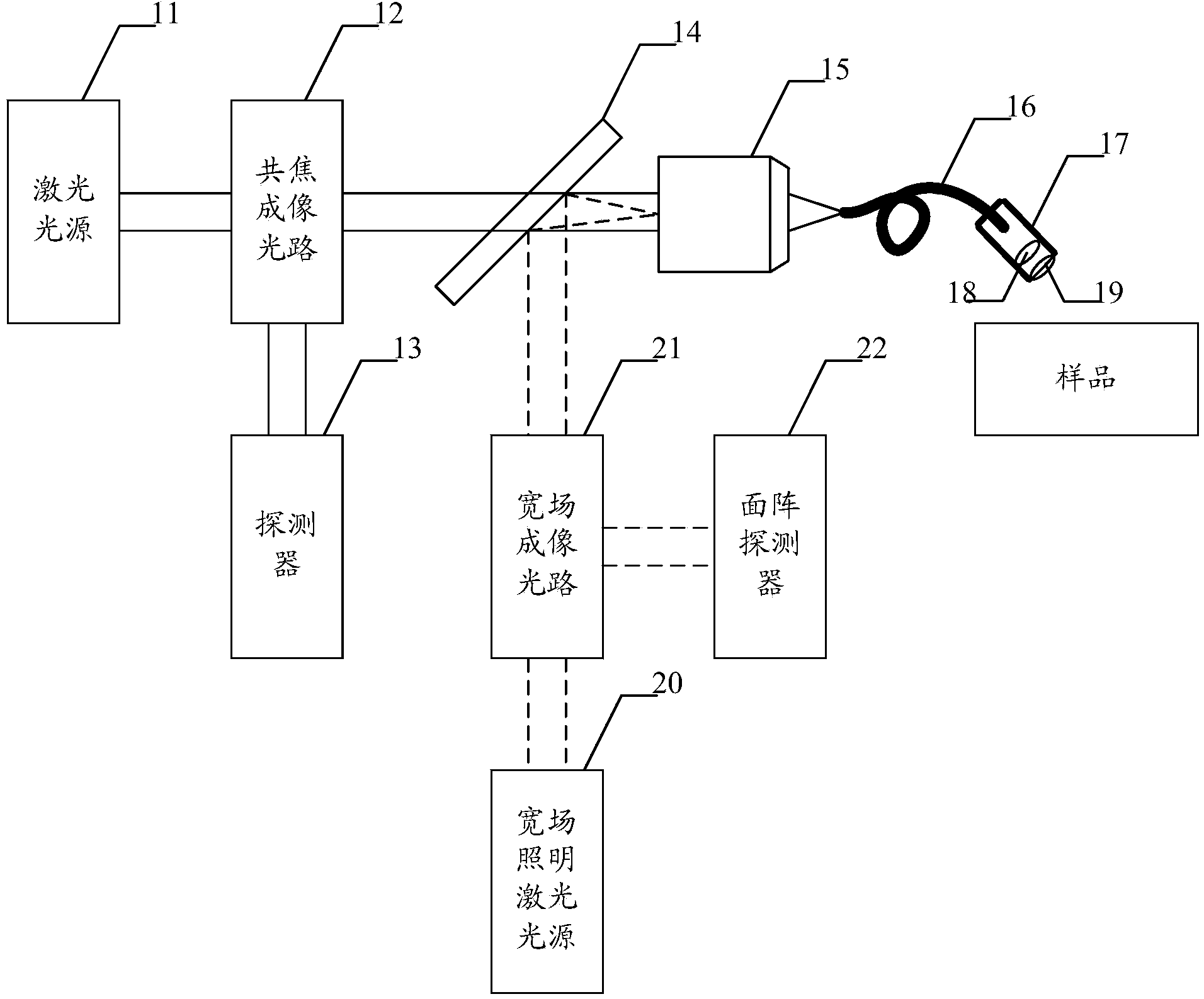
[0004] The purpose of the present invention is to provide a fluorescent confocal microscopic imaging system, which aims to solve the problem that the existing fluorescent confocal microscopic imaging system does not have the ability to quickly and accurately find the target, and needs to use laser point-by-point large-scale scanning, so that the three-dimensional tomography of the target sample can be obtained. The image takes a long time, and it is difficult to accurately image the target object, which makes the system cost high and is not conducive to industrialization.

Method used


Image

  • Laser scanning fluorescence confocal microscopic endoscopic imaging system
  • Laser scanning fluorescence confocal microscopic endoscopic imaging system
  • Laser scanning fluorescence confocal microscopic endoscopic imaging system

Examples


Embodiment Construction

[0022] In order to make the object, technical solution and advantages of the present invention clearer, the present invention will be further described in detail below in conjunction with the accompanying drawings and embodiments. It should be understood that the specific embodiments described here are only used to explain the present invention, not to limit the present invention.

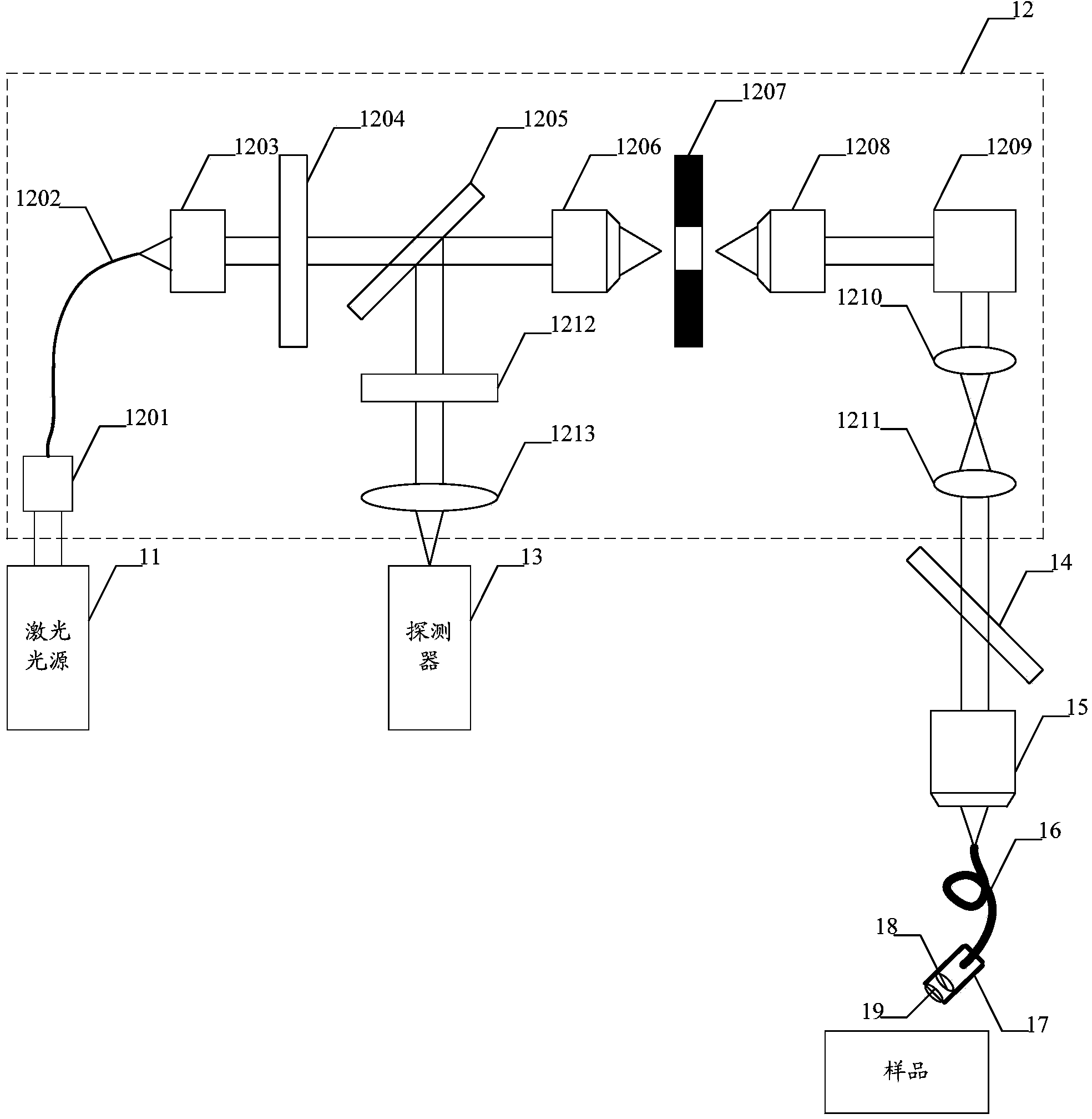
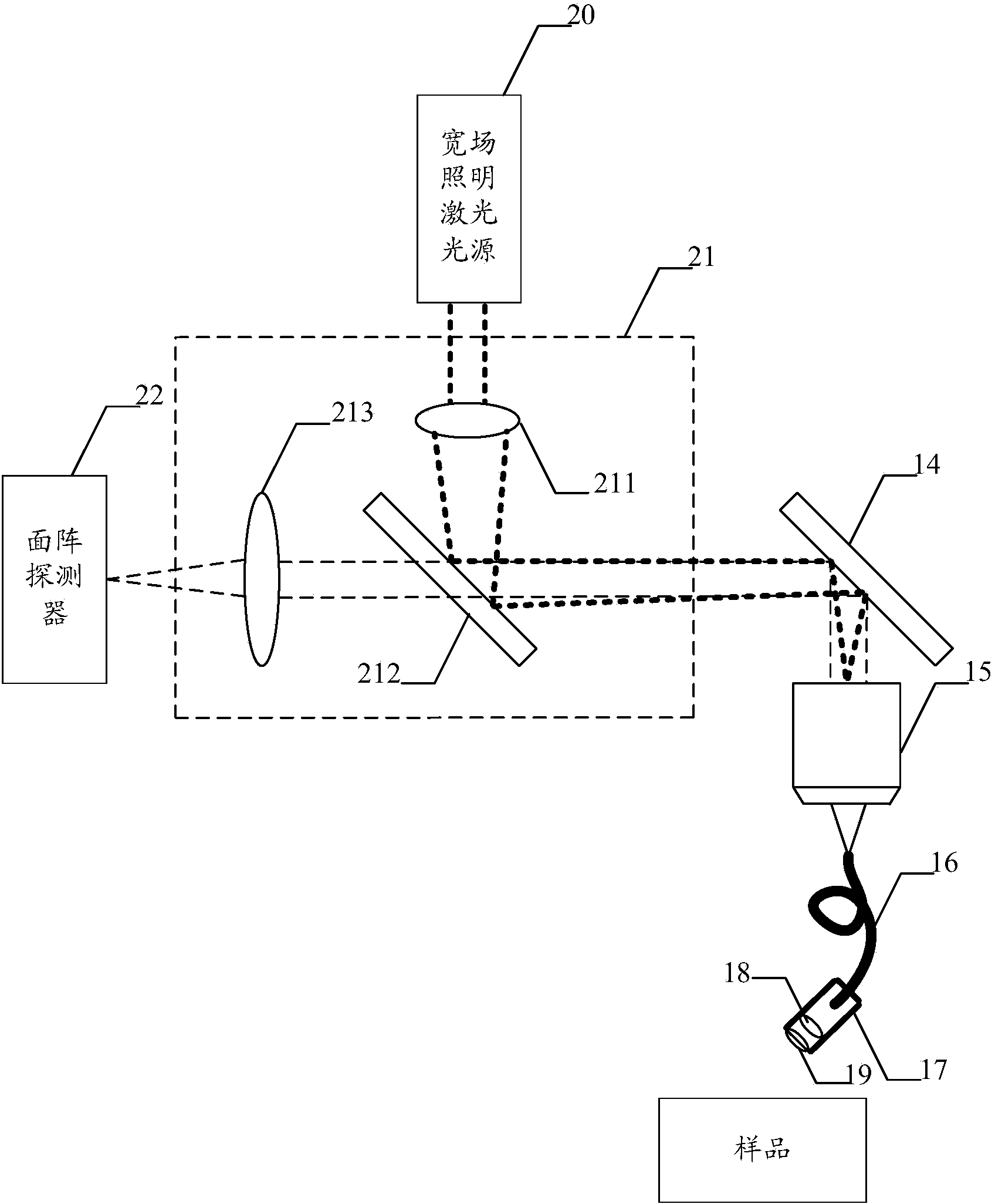
[0023] The fluorescent confocal microscopy imaging system provided by the present invention is based on the existing laser light source and confocal imaging optical path that can obtain three-dimensional tomographic images of samples, and additionally adds a wide-field imaging optical path that can obtain wide-field images and a wide-field illumination laser light source. , and an area array detector, and add optical devices used in conjunction with the confocal imaging optical path and the wide-field imaging optical path, so as to realize the simultaneous display of wide-field images and three-dime...


PUM


Abstract

The invention belongs to the field of three-dimensional microscopic imaging, and provides a fluorescence confocal microscopic imaging system. The system is additionally provided with a wide field imaging light path for obtaining a wide field image, a wide field illumination laser light source and an area-array detector on the basis of a conventional laser light source and a conventional confocal imaging light path which can be used for obtaining a three-dimensional tomographic image of a sample, and optical devices matched with the confocal imaging light path and the wide field imaging light path are also additionally arranged to realize the simultaneous display of the wide field image and the three-dimensional tomographic image. A wide field imaging mode can be used for obtaining the wide field image, so that the rapid search on a target to be observed on the sample is realized, and after the target to be observed is locked, a three-dimensional tomographic imaging mode is used for obtaining a high-accuracy three-dimensional tomographic image of the target to be observed, and simultaneously obtaining a wide field surface image of the sample. Compared with the prior art, the system has the advantages that the problem of long scanning time caused by point-by-point scanning of the sample is solved; in addition, the system is simple in structure, the system cost is lowered, and the industrial application of the system is facilitated.

Description

technical field [0001] The invention belongs to the field of three-dimensional microscopic imaging, in particular to a fluorescent confocal microscopic imaging system. Background technique [0002] The confocal microscopy imaging system is a system that can realize optical tomography imaging of biological samples, etc. In the confocal microscopy imaging system, the light emitted by the point light source is focused to the sample surface by the first objective lens, and then reflected or transmitted by the sample, and then focused to the detector by the condenser lens. The point light source, the object, and the small hole in front of the detector are mutually conjugate. The scanning mechanism scans the sample on the xy plane to obtain a two-dimensional image of the sample, and then obtains the scanned images of multiple layers of the sample by axial scanning, and the images of each layer can be reconstructed after image processing. Resolution 3D tomographic images. [000...

Claims


Application Information

Patent Timeline
IPC IPC(8): G01N21/64G01N21/01
Inventor 邵永红金春平
Owner JIANGSU TIANNING PHOTON TECH
Learn More