
Antigen-removing biological bone and preparation method thereof

A deantigen and biological technology, applied in the field of biomaterials in tissue engineering, can solve the problems of inability to apply orthopedic defect repair, inability to effectively remove antigens, poor compressive performance of bone materials, etc., and achieve good capillary adsorption and retention of mechanical properties Features, the effect of strong pressure resistance

Active Publication Date: 2015-05-27
SHAANXI BIO REGENERATIVE MEDICINE CO LTD
View PDF4 Cites 0 Cited by
  • Summary
  • Abstract
  • Description
  • Claims
  • Application Information

AI Technical Summary

Problems solved by technology

High-temperature calcination can completely remove antigens and organic components, but due to the complete removal of organic matter, the bone material has poor compressive properties, only has osteoconductivity, and has lost its inductivity, so it cannot be used in orthopedic defect repair; and using chemical reagents or Enzyme treatment, for xenograft bone with high antigenicity, cannot effectively remove the antigen, and there is a risk of immune rejection after implantation

Method used


Image

  • Antigen-removing biological bone and preparation method thereof
  • Antigen-removing biological bone and preparation method thereof
  • Antigen-removing biological bone and preparation method thereof

Examples


Embodiment 1

[0037] Embodiment 1: Preparation of deantigenated pork bone

[0038] Step 1. Bone block pretreatment: Take the metaphysis of pig femur and cut it into ≤1cm 3 After the small pieces, wash them with purified water to wash away excess bone marrow and blood, and then use a pulverizer to crush the bone pieces to 5mm-10mm.

[0039] Step 2. Partial deproteinization treatment: Put the bone particles obtained in Step 1 in a mixed solution of 0.5% SDS, 2% sodium hydroxide and 3% hydrogen peroxide; soak at 37°C for 1 hour, and then ultrasonically treat at 40KHz for 3 minutes After the bone particles are fully absorbed by the solution, they are shaken for 2 hours and washed with purified water until neutral. Taking this as a cycle, a total of 4 cycles were treated and then washed with purified water.

[0040] Step 3. Irradiation treatment: use 25-30KGy dose of Co 60 Process the bone particles obtained in step two.

[0041] Step 4. Organic matter removal: The bone particles obtained in...

Embodiment 2

[0045] Embodiment 2: Preparation of deantigenated bovine bone

[0046] Step 1. Pretreatment of bone fragments: cutting bovine femur or porcine femoral metaphysis into bone fragments, washing with purified water until there is no bone marrow and blood; crushing to a size of 5 mm to 10 mm.

[0047] Step 2. Partial deproteinization treatment: Put the bone particles obtained in Step 1 in a mixed solution of 2% Triton100-X, 1% sodium hydroxide and 2% hydrogen peroxide; soak at 37°C for 0.5 hours, and then undergo ultrasonic treatment at 40KHz After 3 minutes, the bone particles are fully absorbed by the solution, shaken for 1.5 hours, and washed with purified water until neutral. Take this as a cycle, and after a total of 4 cycles of treatment, the purified water is washed to neutral.

[0048] Step 3. Irradiation treatment: use 20KGy Co 60 The bone particles obtained in the second step are irradiated.

[0049] Step 4. Removal of organic matter: The bone particles obtained in Ste...

Embodiment 3

[0054] This embodiment is basically the same as Embodiment 1, the difference is:

[0055] Step 2 Partial deproteinization treatment: Put the bone particles obtained in Step 1 in a mixed solution of 1% Triton X-100, 2% sodium hydroxide and 3% hydrogen peroxide; soak at 37°C for 1 hour, and then undergo ultrasonic treatment at 40KHz After 3 minutes, after the bone particles have fully absorbed the solution, shake it for 1.5 hours, and wash it with purified water until neutral.


PUM

PropertyMeasurementUnit
particle diameteraaaaaaaaaa
pore sizeaaaaaaaaaa
crystallinityaaaaaaaaaa

Abstract

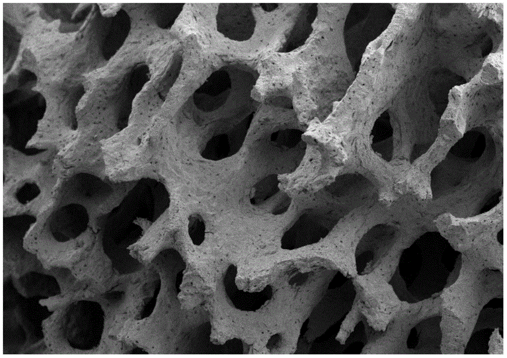
The invention relates to a preparation method of an antigen-removing biological bone. The preparation method comprises bone block pretreatment, partial deproteinization treatment, irradiation treatment, organic matter removal, EDC / NHS crosslinking and drying sterilization. Bone particles are treated by irradiation to make proteins degenerated, then a mixed solution of a surface active agent and hydrogen peroxide is used for treating the irradiated bone particles, followed by a hypochlorite solution and a hydrogen peroxide solution are used for treating, and the deproteinized biological bone is obtained after ethylene oxide sterilization. The prepared bone material has a three-dimensional porous structure, has good capillary adsorption, can adsorb osteoblasts and protein factors after being implanted in bodies, and induces new bone formation; the bone material allows antigen components to be effectively removed, retains a part of collagen, and has better biocompatibility; and the bone material allows hydroxy apatite to have the crystallization degree of lower than 15% and exist in a weak crystallization morphology, has no other crystal phases, allows the carbonate content to be 4%-6%, has relatively high mechanical strength and good degradation, and is suitable for a repairing operation material for non load-bearing part bone coloboma.

Description

technical field [0001] The invention belongs to the biological material technology of tissue engineering, and specifically relates to a deantigenated biological bone and a preparation method thereof. Background technique [0002] Bone grafting has been used in clinical orthopedics for many years. As the gold standard of bone grafting, autologous bone grafting has a good healing effect in clinical practice. The autologous bone is mostly taken from the patient's fibula or hip bone, and the amount of bone taken is limited, and the patient suffers from the pain of a second operation, so the application of autologous bone transplantation is limited. [0003] In order to solve the problem of bone transplantation, artificial bone, allogeneic bone, xenogeneic allogeneic bone and other materials are used instead of autologous bone transplantation for clinical treatment. Artificial bone materials are mostly hydroxyapatite crystals calcined at a high temperature above 1000°C or β-tric...

Claims


Application Information

Patent Timeline
Patent Type & Authority Patents(China)
IPC IPC(8): A61L27/36A61L27/24A61L27/56
Inventor 耿阳雪
Owner SHAANXI BIO REGENERATIVE MEDICINE CO LTD
Learn More