
Construction method of tissue engineering epidermis model

A tissue engineering and construction method technology, applied in tissue culture, biochemical equipment and methods, medical science, etc., can solve problems such as detection fluid leakage, achieve good mechanical properties, ensure mechanical strength, and good adhesion effects

Inactive Publication Date: 2015-03-11
SANITARY EQUIP INST ACAD OF MILITARY MEDICAL SCI PLA +1
View PDF7 Cites 0 Cited by
  • Summary
  • Abstract
  • Description
  • Claims
  • Application Information

AI Technical Summary

Problems solved by technology

[0006] In order to solve the shortcomings of the existing skin models, such as easy shrinkage and leakage of detection fluid at the edge during the culture process, the present invention provides a method for constructing a tissue engineered epidermal model

Method used

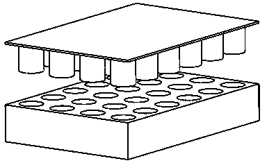

Image

  • Construction method of tissue engineering epidermis model
  • Construction method of tissue engineering epidermis model
  • Construction method of tissue engineering epidermis model

Examples


Embodiment 1

[0029] Polypropylene melt-blown non-woven fabric, hot-pressed at 140°C into a disc shape.

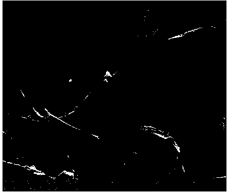
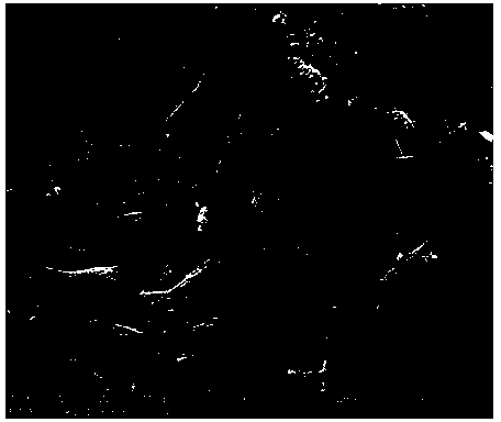
[0030] Commercially available mulberry silk, immersed in 60°C Na 2 CO 3 degumming in aqueous solution, Na 2 CO 3 The concentration of the aqueous solution is 0.2%, 30 minutes each time, 3 times in total, and dried at 70°C after washing with water. Dissolve in a ternary solution of CaCl2, ethanol, and water at 60°C (molar ratio 1:2:8), the dissolution time is 2 hours, put it into a dialysis bag, and dialyze under running water. A silk fibroin solution with a concentration of 3% was obtained. Soak the disc-shaped non-woven fabric in the silk fibroin solution, take it out, dry it in vacuum, and post-treat it with absolute ethanol, with 10 4 / ml Hacat cells were inoculated, cultured on the air-liquid surface, and the tissue engineering epidermal model was constructed. figure 2 is the electron micrograph of the blank scaffold without seeding cells, image 3 It is an electron microgra...

Embodiment 2

[0032] Polyethylene and polypropylene two-component melt-blown non-woven fabric, hot-pressed at 130°C into a disc shape.

[0033] Commercially available mulberry silk is immersed in a neutral soap solution at 90-100°C for degumming, washed with water and then dried at 70°C. Dissolved in CaCl at 95°C 2In the aqueous solution, the dissolution time is 10 minutes, put into a dialysis bag, and dialyze under running water washing. A silk fibroin solution with a concentration of 6% was obtained. Soak the disc-shaped non-woven fabric in the silk fibroin solution, take it out, dry it in vacuum, and post-treat it with absolute ethanol, with 10 4 / ml Hacat cells were inoculated, cultured on the air-liquid surface, and the tissue engineering epidermal model was constructed.

Embodiment 3

[0035] Polyamide spun-bonded non-woven fabric, heat-pressed at 200°C into disc shape.

[0036] Commercially available mulberry silk was degummed by immersing in deionized water under high temperature and pressure, washed with water and dried at 70°C. Dissolved in CaCl at 70°C 2 , ethanol, and water in a ternary solution (molar ratio 1:2:8), the dissolution time is 4 hours, put into a dialysis bag, and dialyze under running water. A silk fibroin solution with a concentration of 9% was obtained. Soak the disc-shaped non-woven fabric in the silk fibroin solution, take it out, dry it in vacuum, and post-treat it with absolute ethanol, with 10 5 / ml Hacat cells were inoculated, cultured on the air-liquid surface, and the tissue engineering epidermal model was constructed.


PUM

PropertyMeasurementUnit
diameteraaaaaaaaaa

Abstract

The invention discloses a construction method of a tissue engineering epidermis model. The construction method comprises the following steps of: A. making a thermoplastic non-woven fabric into a disc-shaped support by using a hot-pressing method; B. by taking mulberry silks as raw materials, degumming, dissolving, dialyzing and concentrating so as to prepare a silk fibroin solution; C. soaking the disc-shaped non-woven fabric support by using the prepared silk fibroin solution, wherein the concentration of the silk fibroin solution is 2 to 10%, and then carrying out vacuum drying and absolute ethyl alcohol aftertreatment; D. inoculating a human epidermis cell strain Hacat, wherein the inoculum density of the Hacat cell is 10<4>-10<5> / ml, carrying out gas-liquid surface cultivation, and proliferating so as to form a lamellar structure, thus constructing the tissue engineering epidermis model. The construction method can be used for solving the shortcoming of the conventional tissue engineering epidermis model that a support is prone to shrink in a cell cultivation process; the edge of the non-woven fabric is fused and bonded by the hot-pressing of the non-woven fabric, so that the problem that the side surface of the constructed non-woven fabric leaks in detection is avoided.

Description

technical field [0001] The invention belongs to the field of tissue engineering in biomedical engineering, and more specifically relates to a method for constructing a tissue engineering epidermis model. Background technique [0002] At home and abroad, instead of animals for skin irritation experiments, tissue-engineered skin models are used. Currently, the alternative skin irritation experimental models recommended by the European Union are the EpiDermTM produced in the United States and the EpiSkin model produced in France. Most of the models use collagen gel as the scaffold for tissue engineering skin. The main disadvantages are that after the cells are seeded on the surface of the gel, the cell / collagen gel complex is easy to shrink during the culture process, and the cell density changes greatly. The area of ​​tissue engineered epidermis also changes, and these deficiencies lead to large errors in the detection of such tissue engineered epidermis. In addition, after e...

Claims


Application Information

Patent Timeline
Patent Type & Authority Patents(China)
IPC IPC(8): C12N5/00A61L27/22A61L27/60
Inventor 李瑞欣张西正卢涛徐成侍才洪郭勇李昊
Owner SANITARY EQUIP INST ACAD OF MILITARY MEDICAL SCI PLA
Learn More