Device for monitoring intraocular pressure
A technology of intraocular pressure and equipment, applied in the direction of tonometer, fluid pressure measurement using capacitance change, glasses/goggles, etc., which can solve the problems of heavy wearing, uncomfortable, and regardless of corneal microstructure
- Summary
- Abstract
- Description
- Claims
- Application Information
AI Technical Summary
Problems solved by technology
Method used
Image
Examples
example 1
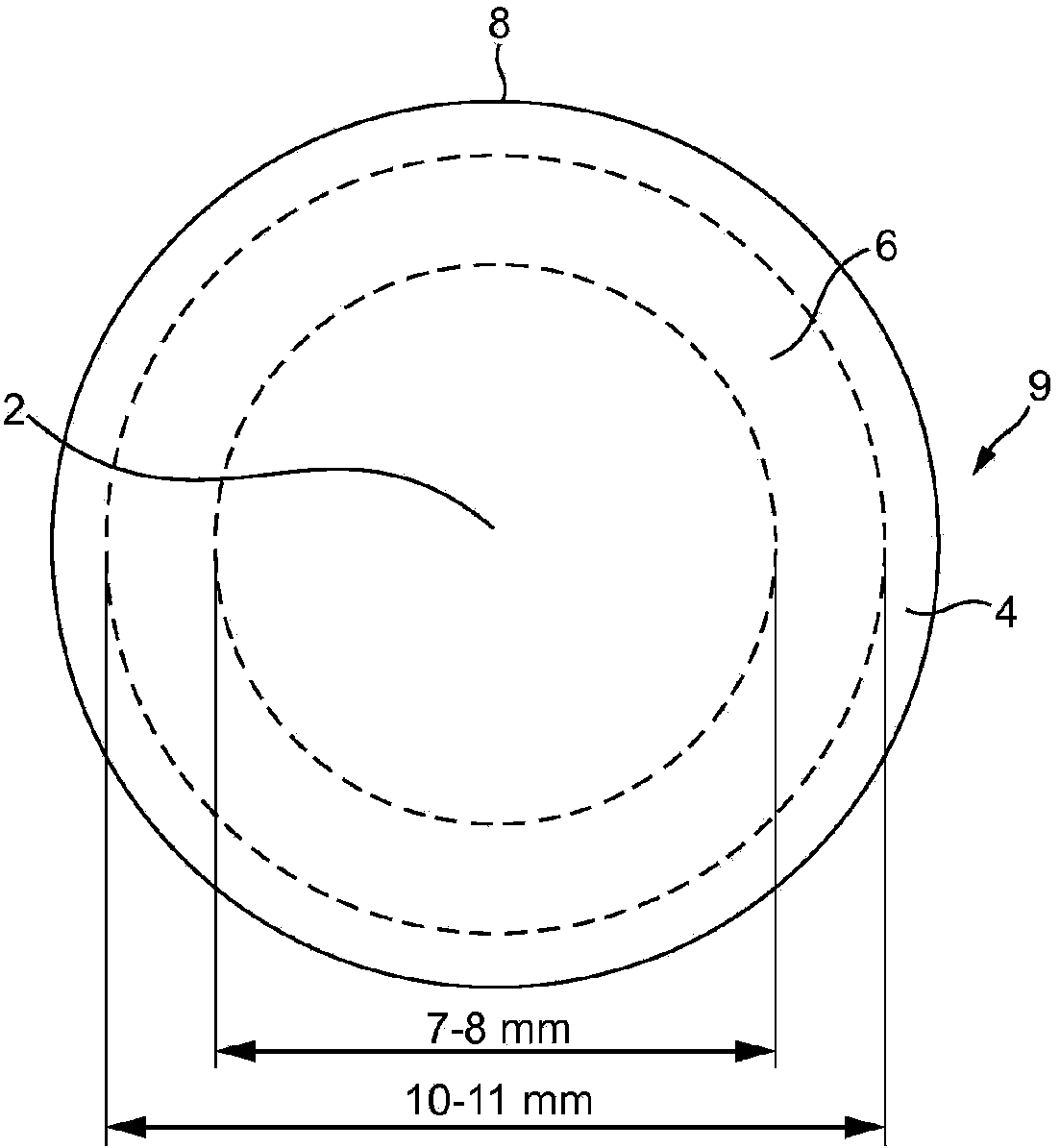
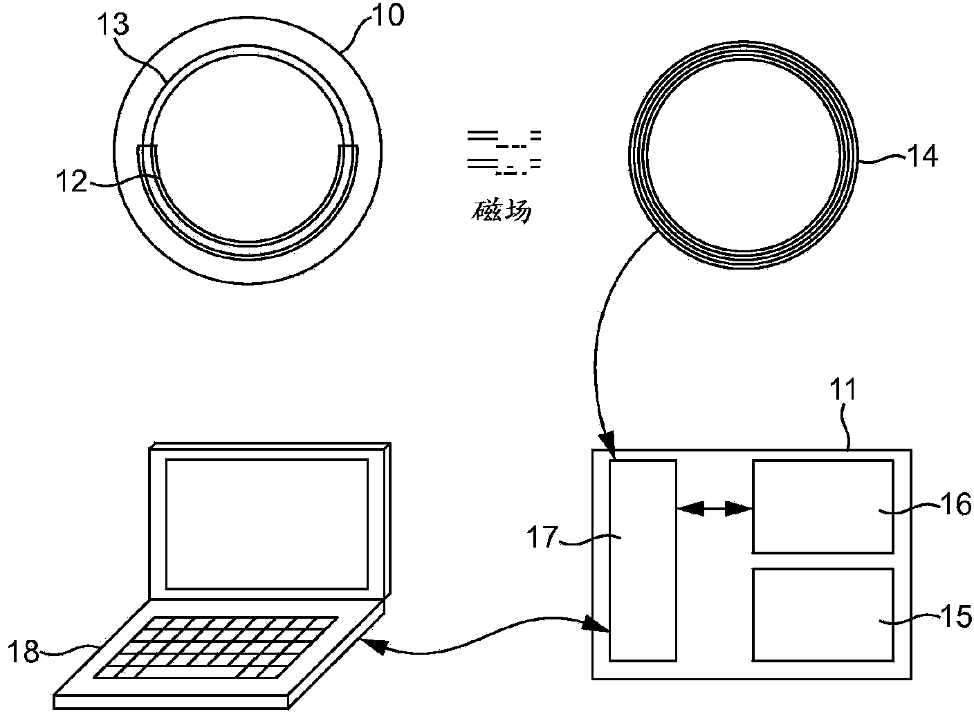
[0106] refer to figure 2 , one embodiment of the device of the present invention comprises two main components: a soft contact lens 10 and an external instrument 11 . Although the dimensions may vary, in this embodiment the contact lens has a diameter of 14-15 mm and a thickness in the range between 120 μm (along the edges) and 280 μm (in the central region). which is composed of a Young's modulus E of 0.30MPa and more than 60×10 -11 cm.mLO 2 / s.Ml.mmHg oxygen transmission rate, soft silicone hydrogel material, which ensures comfortable use over a period of more than 24 hours, and can be adapted to corneas with different sizes and curvatures. Has a 6.0mm central area without any obstruction to achieve a clear view during use. The lens is worn in a similar manner to a normal contact lens based on daily or prolonged wear.
[0107] The contact lens has a circumferential pressure transducer 12 designed to detect small changes in intraocular pressure (IOP) and transmit it wire...
example 2
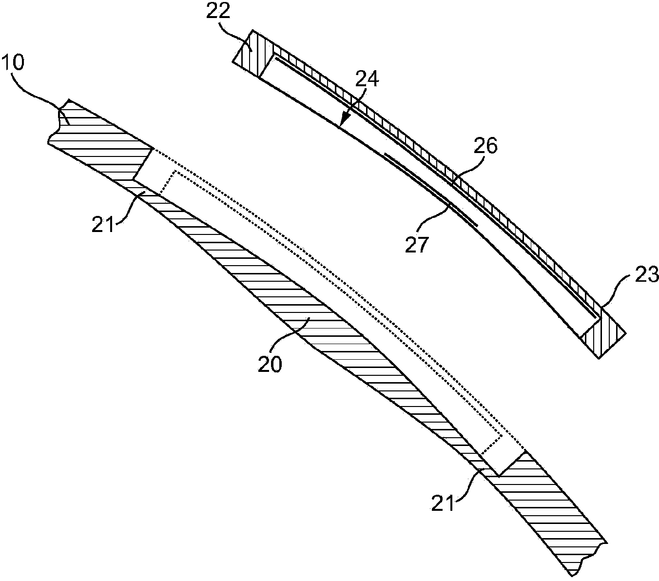
[0110] The following will refer to image 3 and 4 , describing the details of the pressure sensor design. This pressure sensor has been found to produce a stronger IOP signal than previous designs, and is more reliable and stable in its operation in response to eye movement and tear film quality. The design is based on using eyelid pressure to indent the cornea and correlating the resulting reactive deformation of the contact lens to the intraocular pressure (IOP). The design involved modifying the soft contact lens 10 within a typical dimensional area with a 7 mm internal diameter and a 2.5 mm width. like image 3 As shown, the profile in this area is formed with a protruding portion 20 whose peripheral area 21 has been weakened (thinned). The space above the protrusion is covered with a relatively rigid bridge 22 formed by a ring of relatively rigid material having a modulus of elasticity typically around 1 GPa; and a top surface 23 which matches the front profile of the...
example 3
[0121] now refer to Figure 6 a and 6b, The deformation of the diaphragm 24 is measured using a capacitor system comprising two thin metal films 26, 27 fixed to the inner surface of the bridge element and the diaphragm, respectively. When the diaphragm deforms, the distance between the two metal films changes, resulting in a change in the capacitance of the capacitor formed by the two films. This in turn affects the resonant frequency of the oscillator formed by the combination of the capacitance of the capacitor and the inductance of the induction coil. To power the circuit of capacitors and record the resonant frequency value, a magnetic field is formed between an induction coil 28 embedded on the outer surface of the bridge element 22 and an excitation coil 29 placed externally close to the eye (for example on a pair of eyeglasses 30) and the The magnetic field surrounds the induction coil 28 and the excitation coil 29 ( Figure 7). By energizing the field coil with an a...
PUM
 Abstract
Description
Claims
Application Information
 - R&D
- Intellectual Property
- Life Sciences
- Materials
- Tech Scout
- Unparalleled Data Quality
- Higher Quality Content
- 60% Fewer Hallucinations
Browse by: Latest US Patents, China's latest patents, Technical Efficacy Thesaurus, Application Domain, Technology Topic, Popular Technical Reports.
© 2025 PatSnap. All rights reserved.Legal|Privacy policy|Modern Slavery Act Transparency Statement|Sitemap|About US| Contact US: help@patsnap.com


