
Preparation method of polymer based three-dimensional (3D) biochip

A three-dimensional biological and polymer technology, applied in the direction of combinatorial chemistry, library creation, chemical library, etc., can solve the problems of easy diffusion of microarray points, strong instrument dependence, low fluorescence intensity, etc., to achieve the advantages of activity and stability, Overcoming the effect of being easily destroyed and increasing the fluorescence intensity

Inactive Publication Date: 2014-12-31
BEIJING UNIV OF CHEM TECH
View PDF4 Cites 2 Cited by
  • Summary
  • Abstract
  • Description
  • Claims
  • Application Information

AI Technical Summary

Problems solved by technology

[0004] The biochip technology itself also faces many problems to be solved: (1) The hydrophilicity of the biochip carrier is often not good enough, and the adsorption of biological components is very easy to occur, and the surface is polluted by biological components in advance, which affects the follow-up experiments , and the fluorescent background increases, which is not conducive to medical observation; (2) The two-dimensional structure of the surface determines that the amount of biological components that can be combined is extremely limited, and it is impossible to form a large-capacity biological component container, resulting in low fluorescence intensity, which is not conducive to medical observation ; (3) The production speed of biochips is very slow, which depends on the speed at which microliters to nanoliters of samples are added to the surface of the chip carrier by the traditional spotting machine, which is too dependent on the instrument; (4) The sensitivity of biochips , Reproducibility needs to be further improved; (5) Expensive precision spotting equipment limits the development of biochips to a certain extent; (6) Insufficient sources of biological component samples; etc.
Although nitrocellulose membranes and nylon membranes, which are commonly used in traditional Soμthern blotting, have also been used as chip carriers, and these membrane materials can greatly increase the adsorption capacity of biomolecules due to their porosity, but there are also microarray spots. Disadvantages of diffusion, low array density, background fluorescence increase with detection times, and slow hybridization speed restrict its practical application

Method used


Image

  • Preparation method of polymer based three-dimensional (3D) biochip
  • Preparation method of polymer based three-dimensional (3D) biochip
  • Preparation method of polymer based three-dimensional (3D) biochip

Examples


Embodiment 1

[0023] The LDPE membrane was extracted with acetone (extraction and washing solution) for 72 hours, and dried at room temperature. Cover the BOPP film above the LDPE film, inject 150 μL of isopropylthioxanthone acetone solution (concentration: 0.3M) between the double-layer polymers, and then use two quartz plates to compact the double-layer polymers. Propylthioxanthone acetone solution is evenly distributed between the double-layer polymers to form a sandwich structure, and moved into a high-pressure mercury lamp irradiation device (power 1000W, light intensity 10mW / cm 2 ) at room temperature for 180 seconds. The LDPE film that has introduced dormant groups that can continue to initiate free radical polymerization was extracted with acetone for 12 hours to remove isopropylthioxanthone adsorbed on the surface, and dried in a vacuum oven at 25° C. for 2 hours.

[0024] Drop 150 μL of methoxypolyethylene glycol methacrylate solution (monomer molecular weight 1100, solution conc...

Embodiment 2

[0032] Preparation of UV-Vis Spectrophotometric Standard Curve:

[0033] First prepare 1mg / mL BSA stock solution, and then dilute to different concentrations with PBS solution. A total of 9 groups, the concentrations are 0μg / mL, 100μg / mL, 200μg / mL, 400μg / mL, 500μg / mL, 600μg / mL, 800μg / mL, 900μg / mL, 1000μg / mL. The solution with a concentration of 0 μg / mL is the calibration zero solution.

[0034] After the ultraviolet-visible spectrometer is calibrated and reset to zero, take solutions of different concentrations, put 1mL in a cuvette, and scan the absorption at 278nm at a fixed point, that is, A278. The results were repeated 3 times and the average value was taken.

[0035] Use the obtained A278 value to plot with origin and fit it to obtain a standard curve A=(6.31475E-4)c – 0.01853.

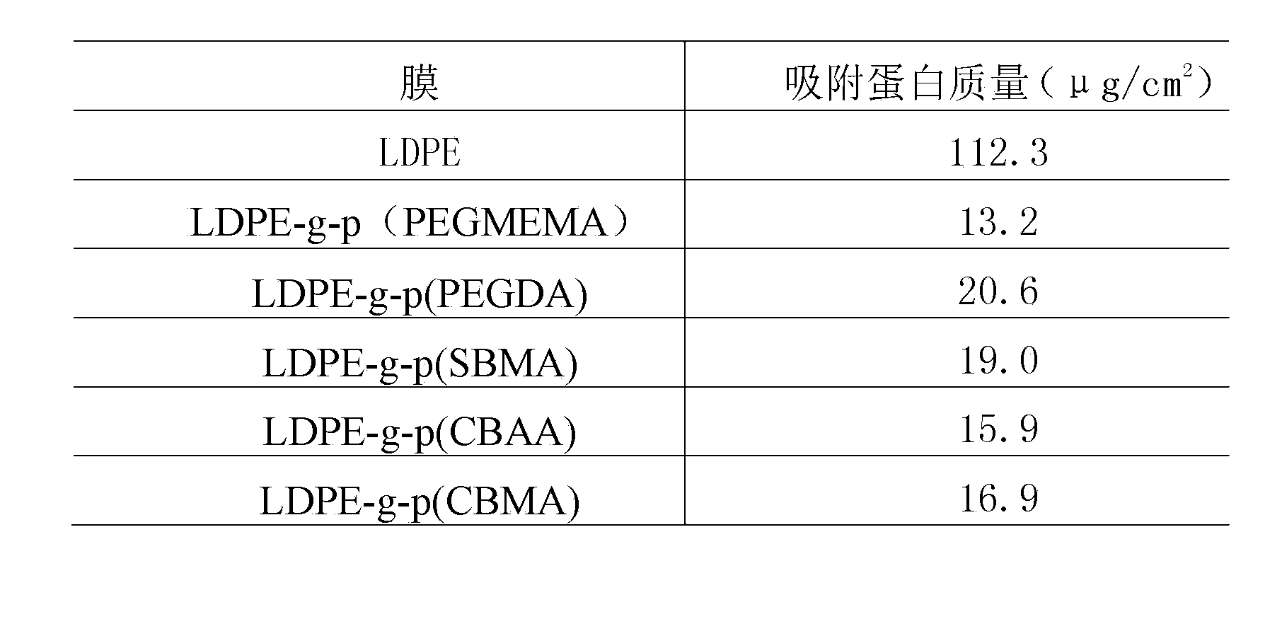
[0036] Anti-biofouling performance test before and after grafting of different anti-biofouling monomers:

[0037] Respectively set the size to 9cm 2 1 sheet of LDPE (No. 1), 1 sheet of LDPE...

Embodiment 3

[0045] The PET film was extracted with acetone (extraction and washing solution) for 72 hours, and dried in a vacuum oven at 25°C for 2 hours. Cover the BOPP film above the PET film, inject 150 μL of benzophenone in acetone solution (concentration: 0.3M) between the double-layer polymers, and then use two quartz plates to compact the double-layer polymers, benzophenone The acetone solution is evenly distributed between the double-layer polymers to form a sandwich structure, and moved into the high-pressure mercury lamp irradiation device (power 1000W, light intensity 16mW / cm 2 ) at room temperature for 180 seconds. The PET film into which the polymerized dormant groups had been introduced was extracted with acetone for 12 hours to remove benzophenone adsorbed on the surface, and dried in a vacuum oven at 25° C. for 2 hours.

[0046] Drop 150 μL of methoxypolyethylene glycol methacrylate solution (monomer molecular weight 1100, solution concentration 0.3M) between the BOPP fil...


PUM

PropertyMeasurementUnit
areaaaaaaaaaaa
thicknessaaaaaaaaaa
diameteraaaaaaaaaa

Abstract

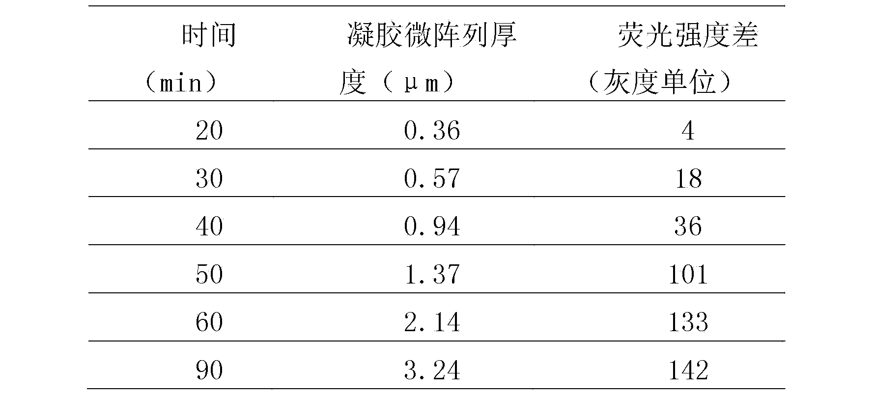
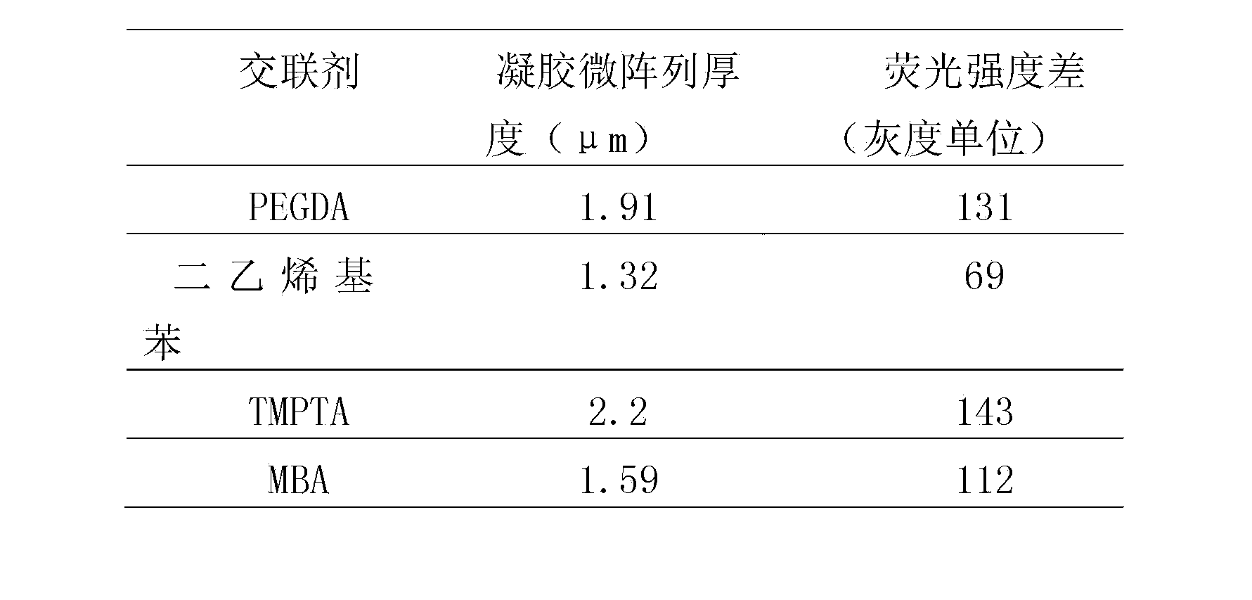
The invention discloses a preparation method of a polymer based three-dimensional (3D) biochip and belongs to the technical field of biochip preparation. The invention discloses a method for preparing the 3D biochip with controllable ingredient content by means of surface activity grafting polymerization technology. The method comprises the following steps of: taking a polymer membrane as a substrate, coupling a semipinacol dormant group on the membrane surface by means of UV (Ultraviolet) radiation, grafting a monomer with biological contamination resistance on the membrane surface by means of visible light / ultraviolet to endow the surface with biological contamination resistance, at last again initiating a grafting monomer mixed solution using a photographic mask under the visible light / ultraviolet to finally obtain the surface functional micro-gel array with controllable thickness, wherein the monomer mixed solution comprises a functional group capable of fixing a biological molecule. The novel 3D biochip, which is low in cost, environment-friendly, simple and convenient in process, high in array point density, high in biomolecule density and strong in fluorescence signal, can be obtained by combining the functional group on the microarray with biologic components of protein, gene and the like, and therefore the preparation method has excellent application prospect.

Description

technical field [0001] The invention belongs to the technical field of biochip preparation, and in particular relates to a method for preparing a biochip by light-initiating polymer film surface active graft polymerization and covalently immobilizing genes, proteins and other biological components. Background technique [0002] With the successful completion of the Human Genome Project, gene chip technology has been further improved and developed. However, many studies have shown that the information of biological function cannot be fully obtained only from the study of genomics. Although the gene chip has been widely used in the field of biomedical research, it can only detect DNA and RNA, and this nucleic acid-based chip detection can only indirectly provide protein information. Therefore, researchers focused their research on protein chips, sugar chips, and cell chips. Protein is the embodiment of life activities, polysaccharides can reflect the level of life activities...

Claims


Application Information

Patent Timeline
Patent Type & Authority Patents(China)
IPC IPC(8): C40B50/00C08J7/18
Inventor 杨万泰林志峰赵长稳马育红朱兴闫煦陈睿超张丽华
Owner BEIJING UNIV OF CHEM TECH
Learn More