
Method for determining microstructure of lipid microsphere/lipid emulsion

A lipid emulsion and microstructure technology, which is applied in the preparation of test samples and material analysis by measuring secondary emissions, can solve the problems of sample damage, low success rate, and poor reproducibility

Inactive Publication Date: 2013-02-27
BEIJING TIDE PHARMA
View PDF5 Cites 2 Cited by
  • Summary
  • Abstract
  • Description
  • Claims
  • Application Information

AI Technical Summary

Problems solved by technology

This method overcomes the defects of low success rate, poor reproducibility, serious damage to the sample, large amount of sample, and inability to truly reflect the microstructure of the sample in the prior art.

Method used


Image

  • Method for determining microstructure of lipid microsphere/lipid emulsion
  • Method for determining microstructure of lipid microsphere/lipid emulsion
  • Method for determining microstructure of lipid microsphere/lipid emulsion

Examples


Embodiment 1

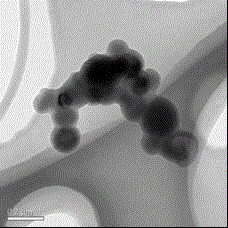
[0033] Sample: alprostadil injection (trade name Kaishi), negative dye: phosphomolybdic acid, dilution ratio: about 2500 times, negative ion generator is Stablo remover from Shimadzu company, drying method is vacuum decompression drying, test The result is as Figure 5 shown.

Embodiment 2

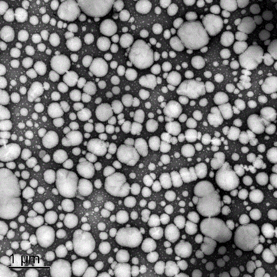
[0035] Sample: Flurbiprofen axetil injection (trade name Kaifen), negative dye: phosphotungstic acid, dilution ratio: about 1000 times, negative ion generator is ANTIST-KIT-UN antistatic device from Mettler, Germany, dry The method is vacuum drying under reduced pressure, and the test results are as follows: Image 6 shown.

Embodiment 3

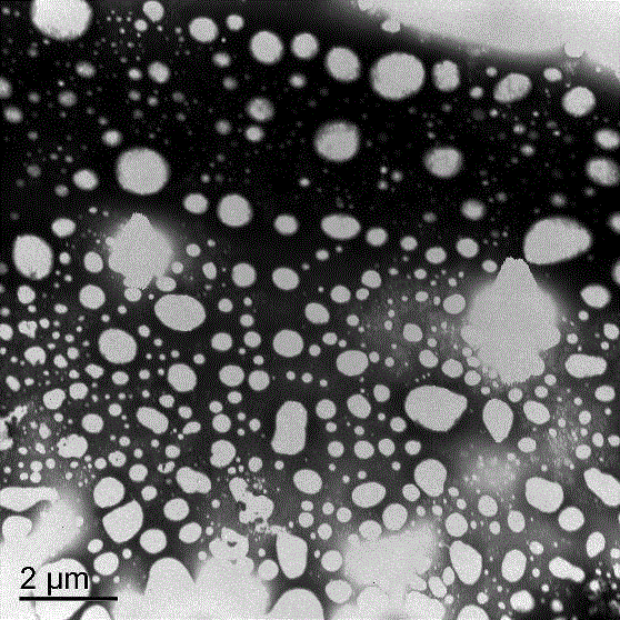
[0037] Sample: fat emulsion injection (trade name Intralipid), negative dye: uranyl acetate, dilution ratio: about 4000 times, negative ion generator is YSTP01 antistatic pen, drying method is infrared lamp drying, the test results are as follows Figure 7 shown.

[0038] Depend on Figure 5 , 6 7. It can be seen that in the three commercially available samples measured by the method of the present invention, the microparticles of the lipid microspheres / lipid emulsion are round, clear, and have good separation, the microscopic morphology is clear, and there is no aggregation and adhesion, and the repeatability of multiple determinations is it is good.


PUM


Abstract

The invention discloses a method for determining the micromorphology of a lipid microsphere / lipid emulsion with a soft shell structure by use of a scanning electron microscope. The method comprises the following steps of: diluting and dispersing a to-be-tested sample, treating the sample by an anion device so as to positively charge the to-be-tested sample or a sample container carrying the to-be-tested sample, and scanning the microstructure of the surface of the sample by the change of electron beam energy, thus obtaining the good-reproducibility, stable and clear microstructure of the lipid microsphere / lipid emulsion, wherein the spectrogram of a scanning TEM (transmission electron microscope) is adopted in the determination method.

Description

technical field [0001] The invention relates to a method for measuring the microstructure of lipid microspheres / lipid emulsions, in particular to a method for measuring a lipid with a soft shell structure by using a negative ion generating device to process a sample to be tested and a sample-loaded metal mesh in a scanning electron microscope. A method for the microscopic morphology of microspheres / lipid emulsions. Background technique [0002] Scanning electron microscope is a relatively modern cell biology research tool invented in 1965. It mainly uses secondary electron signal imaging to observe the surface morphology of the sample, that is, a very narrow electron beam is used to scan the sample, through the interaction between the electron beam and the sample. The action produces various effects, chief among which is the secondary electron emission of the sample. Secondary electrons can generate a magnified topographic image of the sample surface, which is established i...

Claims


Application Information

Patent Timeline
Patent Type & Authority Applications(China)
IPC IPC(8): G01N23/22G01N1/28
Inventor 程栎王伟肖萱刘玉静张伟强杨青松
Owner BEIJING TIDE PHARMA
Learn More