
Sequential separation and mass spectrum identification method of multi-site phosphorylation peptide

An identification method and a phosphorylation technology, which are applied in the direction of material analysis, material analysis, and measurement devices by electromagnetic means, can solve the difficulties in the identification of single phosphorylated peptides and the impossibility of single phosphorylation modification and multi-site phosphorylation modification. Distinguished, easy to break and other problems, to achieve the effect of simple and easy to control the operation process, low cost, and mild reaction conditions

Active Publication Date: 2012-12-12
HUAZHONG NORMAL UNIV
View PDF4 Cites 6 Cited by
  • Summary
  • Abstract
  • Description
  • Claims
  • Application Information

AI Technical Summary

Problems solved by technology

Although existing analysis methods can effectively enrich low-abundance phosphorylated proteins, the coexistence of monophosphorylated peptides and polyphosphorylated peptides not only inhibits each other's signals, but also because phosphorylated modifications are easier to break than polypeptide backbones, multi-site Phosphorylated peptides often generate a series of in-source monophosphate modified peptides, thus making identification of in vivo monophosphorylated peptides difficult
[0003]Existing technologies include IMAC (immobilized metal chelation affinity chromatography), metal oxides, ion exchange and immunoaffinity methods, etc., although they can all enrich phosphoric acid Phosphorylation modified peptides, but can not distinguish between single phosphorylation modification and multi-site phosphorylation modification, only enriched at the same time

Method used


Image

  • Sequential separation and mass spectrum identification method of multi-site phosphorylation peptide
  • Sequential separation and mass spectrum identification method of multi-site phosphorylation peptide
  • Sequential separation and mass spectrum identification method of multi-site phosphorylation peptide

Examples


Embodiment 1

[0060] Sequential Separation of Multi-site Phosphorylated Modified Peptides and Sample Mass Spectrometry Analysis

[0061] 1) Weigh 5~10 mg NiZnFe 2 o 4 Magnetic nano-ferrite material in a centrifuge tube;

[0062] 2), the NiZnFe 2 o 4 The magnetic nano-ferrite material is washed 3 times with a solution containing 50wt% acetonitrile and 0.1wt% TFA, separates the nanoparticles with a magnet, and discards the supernatant;

[0063] 3) Take the NiZnFe obtained in step 2) 2 o 4 The magnetic nano-ferrite particles were washed 3 times with 0.1wt% TFA aqueous solution, and the nanoparticles (NiZnFe 2 o 4 Magnetic beads), discard the supernatant;

[0064] 4) Before analyzing the samples, trypsinize the protein in a 0.1M ammonium bicarbonate solution at 37°C in a water bath for 12 hours;

[0065] 5) Adjust the pH of the enzymolysis solution obtained in step 4) to 1~2 with trifluoroacetic acid (TFA), add acetonitrile and TFA to make it contain 50wt% acetonitrile and 0.1wt% ...

Embodiment 2

[0072] The method of the present invention is used to identify phosphorylated proteins in zebrafish eggs

[0073] 1) Wash zebrafish egg cells with 0.675% saline, add to a glass tissue homogenizer, and mix with cell lysate (the lysate is a buffer solution composed of Tris-HCl and NaCl, and contains detergent 0.1% SDS and 0.5mM enzyme Inhibitor phenylmethylsulfonyl chloride PMSF) mixed.

[0074] 2) Determination of egg protein content by Bradford method;

[0075] 3) The protein disulfide bonds were reduced with dithiothreitol, and derivatized with iodoacetamide, and then trypsinized in a 37°C water bath with a mass ratio of protein:enzyme = 50:1;

[0076] 4) Preparation or purchase of magnetic nano-ferrite materials: NiZnFe 2 o 4 Magnetic beads and Fe 3 o 4 magnetic beads;

[0077] 5), the NiZnFe obtained in step 4) 2 o 4 Magnetic beads and Fe 3 o 4 The magnetic beads were washed 3 times with a washing solution containing 50wt% acetonitrile and 0.1wt% TFA, and then...


PUM


Abstract

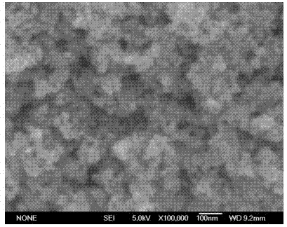
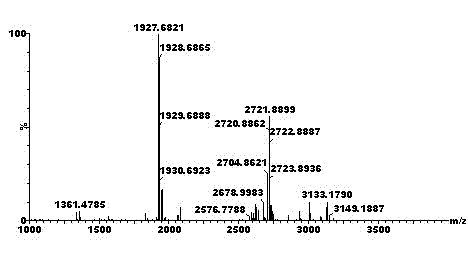
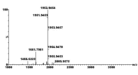
The invention discloses a sequential separation and mass spectrum identification method of multi-site phosphorylation peptide. By using spinel and inverse-spinel type magnetic nano ferrite materials as a separation substrate, the method is used for realizing the fast separation from a complex sample background under the action of an external magnetic field by using the difference of the coordination property between metal ions on an octahedron binding site in the materials and different phosphorylation peptides and the inherent magnetism of the materials. The nano ferrite materials related by the invention can be used for separating a complex polypeptide mixture into non-phosphorylation peptide, mono-phosphorylation peptide and multi-site phosphorylation peptide, so that the signal inhibiting between the molecules of different samples is reduced or eliminated, in-vivo or in-source mono-phosphorylation peptide can be distinguished, and high signal-to-noise ratio and low interference are realized. The method is simple and can be used for effectively enriching low-abundance phosphorylation peptides without using a complex apparatus and realizing the sequential separation and the mass spectrum identification of the multi-site phosphorylation peptide; and the sample analysis and operation are simple without complex sample pre-treatment.

Description

technical field [0001] The invention relates to a method for sequential separation (Sequential Separation) and mass spectrometry identification of multi-site phosphorylated modified peptides. The method utilizes the magnetic nanoparticle of ferrite (Magnetic Nanoparticles of Ferrites, mNOF) material NiZnFe 2 o 4 The differences in the affinity between metal ions and different phosphorylated peptides in the octahedral lattice unit of the middle surface, as well as their inherent magnetic properties, sequentially separate phosphorylated peptides in complex biological samples into single phosphorylated modified peptides and multi-site Phosphorylation modifies the two parts of the peptide, thereby reducing or eliminating the mutual signal inhibition between ions and achieving in vivo (in vivo) or in-source (in the ion source) mass spectrometric determination of monophosphorylated modified polypeptides. Background technique [0002] A large number of proteins in cells not only...

Claims


Application Information

Patent Timeline
IPC IPC(8): G01N27/62
Inventor 钟鸿英肖潇胡雪娇郑石黄璐璐
Owner HUAZHONG NORMAL UNIV
Learn More