
Novel stem cells, method for screening same, kit and application thereof

A technology of stem cells and kits, applied in the fields of new stem cells, their screening, kits and their uses, can solve problems such as low cell viability, affecting cell viability, improper operation, etc.

Inactive Publication Date: 2012-11-28
李福生 +1
View PDF3 Cites 3 Cited by
  • Summary
  • Abstract
  • Description
  • Claims
  • Application Information

AI Technical Summary

Problems solved by technology

Through experiments such as the mouse genetic marker lineage test, combined with the discovered molecular markers, such as Musashi-1, Lgr5, etc., it is possible to identify stem cells (Barker N et al., Nature, 2007.449(7165): 1003-1007; Haegebarth A et al., Am J Pathol, 2009.174(3):715-721), however, their effectiveness remains to be further confirmed
The disadvantage is that the magnetic bead labeling may affect the cell viability, and the enzymatic digestion takes a long time, the cell viability after enrichment is not high, and these isolated cell populations are not of homogeneous type, but are composed of many different differentiated cells. Composition of mixed cell populations in state or proliferative capacity, very impure
This method has been used to screen potential stem / progenitor cell populations from other organs and tissues. The disadvantage is that when SP cells are screened from solid tissues, due to the limited ability to obtain viable cells, when such cells are May affect their ability to remodel after transplantation
[0013] 7) Based on the functional characteristics of stem cells, such as resistance to treatment with drugs or other agents, methods for activating stem cells have been reported, (Gordon MY et al., Leukemia research, 1985.9: 1017-1021; Berardi AC et al., Science, 1995.267: 104 -108; Sharkis SJ et al, Stem ceHs, 1997.15 (Suppl 1): 41-44; Juopperi TA et al, Experimental hematology, 2007.35: 335-341), and use some molecular surface markers to separate these cells, however, the disadvantage , on the one hand, the molecular markers they used are still not very specific, and it is still unclear whether there are progenitor cells of other stages and proliferating cells that originally existed, and it is likely that it is still a heterogeneous cell population ( Berardi AC et al., Science, 1995.267: 104-108), more importantly, no further reliable and generally valid methods for identifying these activated stem cells
[0015] The methods or technologies disclosed above have both advantages and disadvantages. For example, flow cytometry and immunomagnetic bead sorting often screen and isolate stem cells based on special markers on the cell surface, but currently specific cell surface markers are difficult to obtain. Reached a consensus that there is a lack of specific molecular surface markers. In addition, it needs to be completed with the help of professional operating instruments. The separation time is longer and the cost is higher. And through certain treatments, it is equipped with labels that can be recognized by the instrument. This chemical process It will have a certain impact on the cell state. Improper operation may even cause differentiation or lead to cell apoptosis. On the other hand, the sorting efficiency of the instrument is not high, and the long-term sorting process will lead to the decline of cell viability and the change of cell state. , these are not suitable for large-scale screening and enrichment of stem cells for scientific research, clinical use, etc.
[0016] Furthermore, there is a lack of functional evidence that LRCs have the functional characteristics of stem cells, and BrdU is not a cell surface marker (Blanpain et al., Cell, 2004.118(5): 635-648); isolation of stem cells through nuclear morphology requires high expertise and experience , it is prone to identification errors, and the operation process and time are relatively long

Method used


Image

  • Novel stem cells, method for screening same, kit and application thereof
  • Novel stem cells, method for screening same, kit and application thereof
  • Novel stem cells, method for screening same, kit and application thereof

Examples


Embodiment 1

[0141] Example 1 Screening and Identification of New Liver Stem Cells

[0142] The liver tissue is obtained from rodents, humans or other mammals, normal liver tissue, biopsy or isolated liver tissue, and then the tissue is washed with PBS buffer solution for 2 to 3 times, and then the liver tissue is cut into pieces with ophthalmic scissors. 1mm 3 After adding trypsin (concentration: 0.1%~0.3%), digest at 37°C for 10mim~60min, add DMEM / F12K (Hyclone Company, USA) containing 10% fetal bovine serum equal to the volume of trypsin solution to stop Reaction, centrifuge at 800-1000g for 3mim-5min at low temperature, discard the supernatant, add low-temperature pre-cooled PBS containing 2% fetal bovine serum to wash the precipitate for 2-3 times, and filter with a sterile 100-400-mesh sieve to filter Take out the cell suspension, trypan blue staining live cells ≥ 96.7%.

[0143] Inoculate the isolated primary cell population in cell culture flasks or cell culture plates or culture...

Embodiment 2

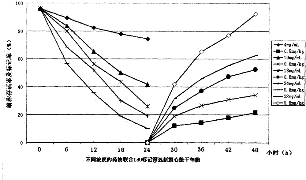
[0152] Example 2 Screening and Identification of New Cardiac Stem Cells

[0153] The heart tissue is obtained from the normal heart tissue of rodents, humans or other mammals, biopsy or isolated fresh heart tissue, and then the tissue is washed with PBS buffer for 2 to 3 times, and then the heart tissue is cut into pieces with ophthalmic scissors. About 0.5~1mm 3 Add collagenase (concentration: 2-10mg / mL) and digest at 37°C for 10mim-30min, add DMEM / F12K containing 10% fetal bovine serum (U.S. Hyclone Company) equal to the volume of collagenase solution to stop For reaction, centrifuge at 900 g for 3 min to 5 min at low temperature, discard the supernatant, add low-temperature pre-cooled PBS containing 2% fetal bovine serum to wash the precipitate for 2 to 3 times, and filter with a sterile 200 to 400 mesh screen to filter out the cell suspension. Solution, trypan blue staining live cells ≥ 95%.

[0154] The cell population was seeded in a 6-well cell culture plate at 37°C, ...

Embodiment 3

[0163] Example 3 Screening of novel cancer stem cells from liver cancer

[0164] Liver cancer tissues were obtained from rodents, humans or other mammals, biopsies, or fresh liver cancer tissues in vitro, and then washed with PBS buffer for 2 to 3 times, and then cut into pieces with ophthalmic scissors. about 1mm 3 Add trypsin (concentration: 0.1% ~ 0.3%) and collagenase (concentration: 1 ~ 10mg / mL) and digest at 37°C for 5mim ~ 60min, add the same volume of enzyme solution containing 10% DMEM / F12K (U.S. Hyclone Company) of fetal bovine serum was used to stop the reaction, and the cell suspension was gently blown 2-3 times, centrifuged at 800-1000g for 3mim-5min at low temperature, discarded the supernatant, and added low-temperature pre-cooled 2% fetal bovine The serum was washed with PBS for two to three times, filtered through a sterile 200-400-mesh sieve to filter out the cell suspension, and trypan blue stained viable cells > 96%.

[0165] Inoculate the isolated primar...


PUM


Abstract

The invention belongs to the technical field of cells and discloses novel adult stem cells, a method for screening the same, a kit and application thereof. Biopsy samples, in vitro tissue samples, and cell line samples of normal tissues and tumors, precancerous tissues and tumors, carcinogenic tissues and metastatic tumors or cancer tissues of rodent, human or other mammals are screened by using chemotherapeutics and cell markers which are combined so as to obtain the stem cell groups. Moreover, the invention also discloses a method for screening and identifying the stem cells from the mixed cell groups, and the kit; and the method and the kit have the advantages of strong specificity, simplicity, convenience, quickness, practicality and effectiveness. The novel stem cells, the method for screening the same and the kit can be applied to clinic treatment, basis and application research, treatment, tissue engineering, medicine screening, and repairing and regeneration of diseased tissues and organs which loose functions well, and have wide prospect.

Description

technical field [0001] The present invention belongs to the field of cell technology, and in particular relates to a novel adult stem cell, its screening method, a kit and its use, and in particular relates to the method of combining chemotherapeutic drugs with cell markers from rodents, humans or other mammals. , tumor / precancerous, neoplastic / cancerous, metastatic / cancerous tissue biopsies, ex vivo tissue samples, stem cells screened from cell line samples, and more specifically, involves pre-treatment of cell populations with chemotherapeutic drugs to remove them Proliferative cell populations, and then screen, enrich and activate the stem cell populations, and then use the markers that can mark the stem cells to identify them and or their progeny. Kits for screening and labeling new adult stem cells, and the application of the screened new adult stem cells in clinical, basic and applied research, treatment, tissue engineering, drug screening, repair and regeneration of dis...

Claims


Application Information

Patent Timeline
IPC IPC(8): C12N5/074C12N5/095C12Q1/04A61L27/38A61K35/12C12Q1/02A61K35/545
Inventor 李福生卢磊磊卢淼淼卢晶晶
Owner 李福生
Learn More