A visual digital ophthalmoscope

An ophthalmoscope and digital technology, applied in the field of ophthalmoscope, can solve the problems of doctors' eye fatigue, small observation field of view, unfavorable diagnosis, etc., and achieve the effect of improving the accuracy rate, easy to carry, and simple structure

Active Publication Date: 2014-10-01
SUZHOU KANGJIE MEDICAL
View PDF4 Cites 0 Cited by
  • Summary
  • Abstract
  • Description
  • Claims
  • Application Information

AI Technical Summary

Problems solved by technology

[0003] 1. During the whole inspection, the eyes are close to the ophthalmoscope, and the close observation is completed, which is easy to cause secondary infection to the patient's eyes;
[0004] 2. The doctor's observation field of view is small, which may easily cause the doctor's eye fatigue and cause misdiagnosis;
[0005] 3. The diagnosis results are not easy to save, and the conclusions drawn simply through the doctor's observation may be biased, which is not conducive to further diagnosis in the later stage

Method used


Image

  • A visual digital ophthalmoscope
  • A visual digital ophthalmoscope
  • A visual digital ophthalmoscope

Examples


Embodiment Construction

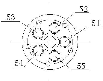
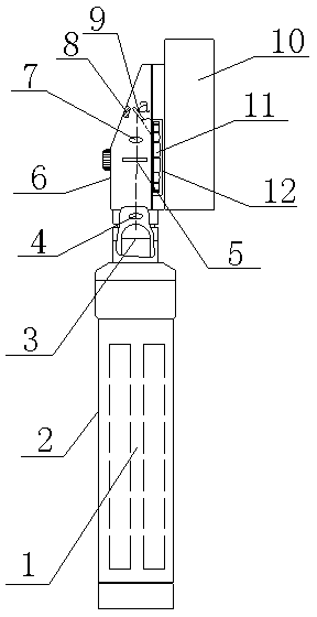
[0017] figure 1 , figure 2 A visual digital ophthalmoscope for checking and recording fundus diseases is shown, which includes a handle 2 and a main housing 6 arranged on the upper part of the handle 2. The main housing 6 is provided with a rotating wheel 11, and the right side of the rotating wheel 11 is The diopter compensation lens 12 is sleeved, and its diopter refers to the ability to express refraction with light intensity. The compensation lens is obtained by introducing diopters of equal size and opposite directions into the undesired toric surface produced by the processing process. The function of the diopter compensation mirror 12 is to gather the light reflected from the fundus 13 of the patient into the digital video camera 10; Shooting and recording; the left side of the main casing 6 is an inclined plane, and an observation hole 8 is opened on the inclined surface. The main casing 6 is sequentially provided with a light bulb 3, a condenser lens 4, an adjustme...


PUM


Abstract

The invention relates to a visual digital ophthalmoscope for checking and recording fundus diseases, which includes a handle and a main casing arranged on the upper part of the handle. A rotating wheel is arranged inside the main casing, and a diopter compensation mirror is sleeved on the right side of the rotating wheel. The right side of the main casing is provided with a digital video camera, and the main casing is sequentially provided with a light bulb, a condenser, an adjusting disc, a projection mirror and a reflector from bottom to top, and the placement position of the reflector is at an angle of 45° to the right side wall of the main casing. The light emitted by the bulb enters the adjustment disc after being converged by the condenser lens, and then the light is coupled out into the projection mirror by the slit in the adjustment disc. The light from the light enters the digital camera through the diopter compensation lens, and the digital camera can shoot, record, record and save the fundus mirror image of the patient to improve the correct rate of diagnosis; the ophthalmoscope with this structure is small in size, simple in structure, easy to carry, and is suitable for going out for medical treatment.

Description

technical field [0001] The invention relates to an ophthalmoscope used for eye fundus disease inspection, in particular to a portable visible digital ophthalmoscope with the functions of inspection, photographing, recording and saving. Background technique [0002] It is usually used clinically to check the fundus lesions of patients, such as retinal detachment, neurological atrophy, cataract, macular hemorrhage, etc., through the large ophthalmoscope used in the hospital for medical examination to determine the location and nature of the lesion. At present, the ophthalmoscope widely used in China is mainly used in hospitals. It is composed of a light source, a condenser, a steering mirror and a variable eyepiece. The internally reflected light observes the condition of the fundus, but there are many disadvantages: [0003] 1. During the whole inspection, the eyes are close to the ophthalmoscope, and the close observation is completed, which is easy to cause secondary infec...

Claims


Application Information

Patent Timeline
Patent Type & Authority Patents(China)
IPC IPC(8): A61B3/12A61B3/14
Inventor 朱永林
Owner SUZHOU KANGJIE MEDICAL
Why Eureka
Try Eureka