
A Real-time Ultrasound Image Simulation Method Based on CT Volume Data

A technology of ultrasound image and simulation method, applied in the field of medical ultrasound training, can solve problems such as poor real-time effect of ultrasound simulation, high distortion rate of ultrasound images, speckle noise of ultrasound images, etc., to achieve increased authenticity, good real-time performance, and low distortion rate Effect

Active Publication Date: 2015-08-19
ARIEMEDI MEDICAL SCI BEIJING CO LTD
View PDF3 Cites 0 Cited by
  • Summary
  • Abstract
  • Description
  • Claims
  • Application Information

AI Technical Summary

Problems solved by technology

[0004] However, when the ultrasonic beam entering the human body is scattered, the scattered echoes with different phases interfere with each other, resulting in speckle noise in the ultrasonic image. Doctors need a wealth of knowledge to accurately identify human tissues and organs and their lesions. On the other hand, by obtaining There are many limitations in real-time hypermap images for ultrasound training and it is impossible to be popularized. Ultrasound image simulation has been proposed as a convenient training method, but the current ultrasound image simulation has several defects:
[0005] 1. Using existing ultrasound images for simulation, when the ultrasound probe moves outside the area, the simulation effect is poor;
[0006] 2. Use FieldII software for simulation, because of the large calculation amount of FieldII, the real-time effect of ultrasonic simulation is very poor;
[0007] 3. Only the reflection coefficient of the edge pixels in the image is calculated, and the distortion rate of the ultrasonic image is high

Method used


Image

  • A Real-time Ultrasound Image Simulation Method Based on CT Volume Data
  • A Real-time Ultrasound Image Simulation Method Based on CT Volume Data
  • A Real-time Ultrasound Image Simulation Method Based on CT Volume Data

Examples


Embodiment Construction

[0026] The advantages and spirit of the present invention can be further understood through the following detailed description of the invention and the accompanying drawings.

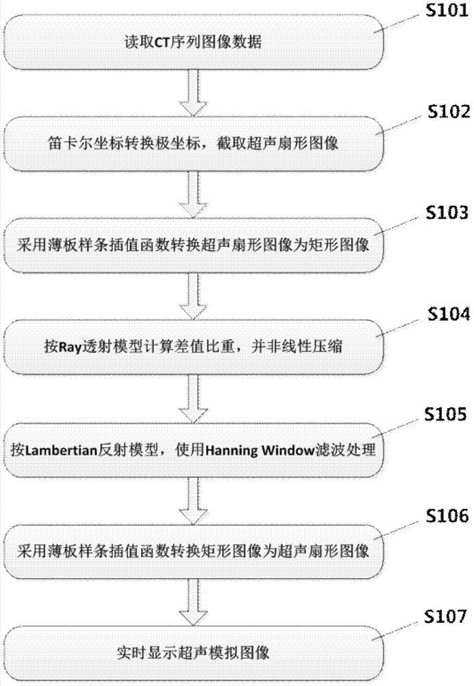
[0027] attached figure 1 To simulate the flow chart, the medical image display and interaction include the following steps:

[0028] Step S101, read CT volume data, the CT volume data can be DICOM serial slice images, or RAW three-dimensional volume data.

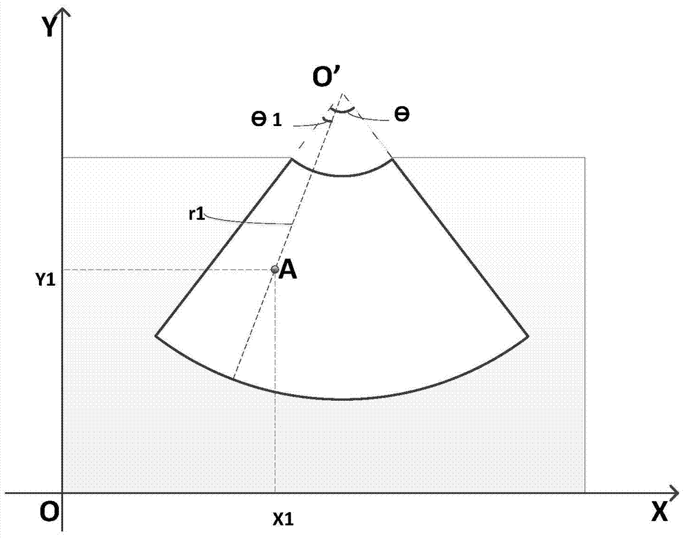
[0029] Step S102, extracting the cross-section of the read CT volume data to obtain 2D image data, and converting Cartesian coordinates to polar coordinates according to the parameters of the actual ultrasound equipment, and intercepting the ultrasound fan-shaped image. The conversion formula is as follows:

[0030] x=x 0 +(r 0 +n·Δr)·sin(m·Δθ+θ 0 )

[0031] y=y 0 +(r 0 +n·Δr)·cos(m·Δθ+θ 0 ) (1)

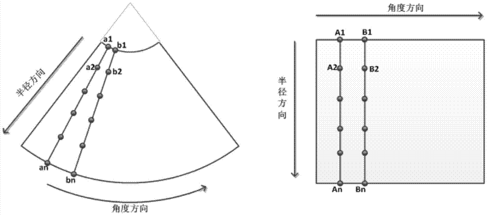
[0032] Step S103, using the thin-plate spline interpolation function to map the ultrasonic fan-shaped image into a rectangular image. Th...


PUM


Abstract

An ultrasonic image needs a doctor to have abundant ultrasonic image recognizing experiences when being used for recognizing and judging human tissue posterior ischic optico-neuropthy, and real-time ultrasonic image simulation provides a convenient tool for ultrasonic training. An ultrasonic image simulating method is used for simulating the ultrasonic image in real time on the basis of CT volume data, carrying out inter-conversion on coordinate positions of pixel points of an ultrasonic sector image and a CT rectangular image by adopting a thin plate spline interpolation function, obtaining a reflectivity of a tissue organ interface according to a Lambertian reflection model by using nonlinear compression conversion through calculating a difference proportion of CT values between the two pixel points of a single Ray by combining the characteristic of a tissue organ edge detection operator and the characteristic of CT image and ultrasonic image noise, and realizing simulation of an ultrasonic probe multi-unit transducer through a Hanning Window function. The ultrasonic image simulation has the advantages of high trueness and strong instantaneity.

Description

technical field [0001] The invention relates to a real-time ultrasound image simulation method based on CT volume data, which is suitable for the field of medical ultrasound training. Background technique [0002] The principle of CT imaging is: use X-ray beams to scan a layer of a certain thickness of the human body, the detector receives the X-rays that pass through this layer, converts them into visible light, converts them from photoelectricity to electrical signals, and then passes through analog / digital signals. The converter (analog / digital converter) converts it into a number and inputs it into a computer for processing. The processing of image formation is like dividing the selected layer into several cuboids with the same volume, which are called voxels. The scanned information is calculated to obtain the X-ray attenuation coefficient or absorption coefficient of each voxel, and then arranged into a matrix, that is, a digital matrix. The digital matrix can be stor...

Claims


Application Information

Patent Timeline
Patent Type & Authority Patents(China)
IPC IPC(8): G09B19/00
Inventor 杨健丛伟建刘越王涌天
Owner ARIEMEDI MEDICAL SCI BEIJING CO LTD
Learn More