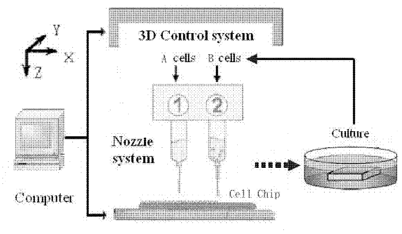
Three-dimensional cell chip based on cell printing and multi-parameter sensing array integrated technology
A cell chip, cell technology, applied in the field of biological information, can solve the problems of limiting real-time detection, cell damage, cell death, etc.
- Summary
- Abstract
- Description
- Claims
- Application Information
AI Technical Summary
Problems solved by technology
Method used
Image
Examples
Embodiment 1
[0056] Embodiment 1 cardiomyocyte culture
[0057] Cardiomyocytes derived from neonatal rats were selected as one of the seed cells forming the basis of the system.
[0058] (1) Take one suckling mouse at a time, put it on a piece of sterile gauze, scrub its chest and abdomen with 70% ethanol, remove the heart and put it into the beaker marked 1. Each dissection should not exceed 1 minute.
[0059] (2) Use a hemostat to remove the connective tissue and blood clots on the heart, then transfer it to the beaker marked 2, wash it again, and transfer it to the beaker marked 3. Use a straw to gently suck out the D-Hanks solution in beaker 3, then add 2ml of 0.25% trypsin solution (37°C), use ophthalmic scissors to cut the heart into sand-sized pieces, add 2ml of trypsin solution, and mix well After digestion at 37°C for 15 minutes.
[0060] (3) After digestion, suck off the supernatant, add 4ml of new trypsin solution, mix well, digest at 37°C for 15 minutes, then discard the sup...
Embodiment 2
[0066] Embodiment 2 adrenal chromaffin cell culture
[0067] (1) PC-12 mouse adrenal pheochromocytoma cell line (ATCC CRL-1721, American Type Culture Collection, American Type Culture Collection Center), with high sugar DMEM (Dulbecco's Modified Eagle's Medium, Sigma) + 10% fetal bovine Serum culture medium, cultured at 37°C carbon dioxide constant temperature culture, generally about three days, the cells reach monolayer confluence, after the cells are confluent, carry out routine subculture;
[0068] (2) Subculture: Absorb the culture medium, add 1 mL of 0.2% trypsin solution, quickly wash the bottle wall and then absorb it, then add 1 mL of 0.2% trypsin solution, incubate at 37°C for about 5 minutes, observe under a microscope, and wait for 80% of the After the cells become round, gently absorb the trypsin solution, and quickly add DMEM medium containing 15% FCS to stop the digestion; blow the wall of the culture flask to make a cell suspension again; adjust the cell concen...
Embodiment 3
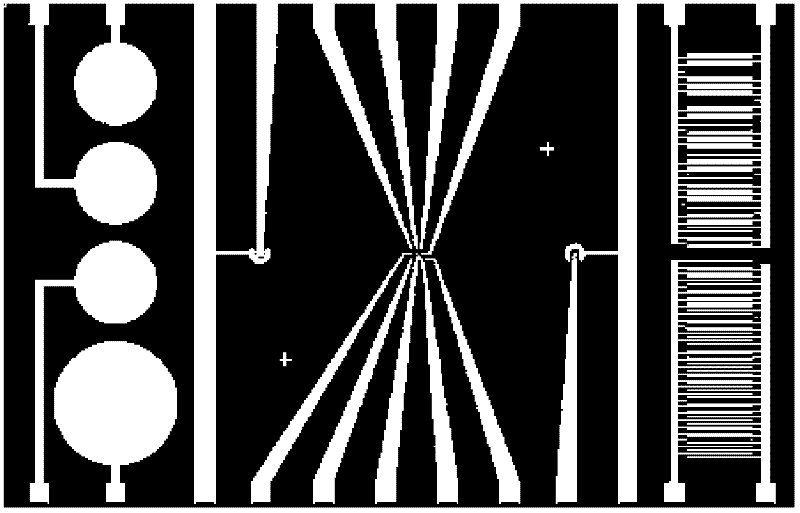
[0070] Embodiment 3 Design and manufacture of multi-sensor integrated chip
[0071] In the processing technology, both the MEA and ECIS units use gold electrodes and the base structure and protective layer structure are the same, so the processing technology can be shared. For specific processing procedures, see image 3 shown in each step. The processing process takes the silicon substrate as an example. The processing process is briefly described as follows:
[0072]First, the insulating layer on the surface of the device is grown and deposited, and a layer of 100nm Si3N4 is deposited on it by chemical vapor deposition (PECVD) process. Then process multiple functional units of MEA and ECIS. On the basis of the previous steps of processing, a layer of SiO2 with a thickness of 600nm is deposited on the chip by PECVD. The metal layer adopts Au / Cr composite layer, and Cr is used to enhance the adhesion between gold and the silicon base layer on which SiO2 is deposited. , and...
PUM
 Abstract
Description
Claims
Application Information
 - R&D
- Intellectual Property
- Life Sciences
- Materials
- Tech Scout
- Unparalleled Data Quality
- Higher Quality Content
- 60% Fewer Hallucinations
Browse by: Latest US Patents, China's latest patents, Technical Efficacy Thesaurus, Application Domain, Technology Topic, Popular Technical Reports.
© 2025 PatSnap. All rights reserved.Legal|Privacy policy|Modern Slavery Act Transparency Statement|Sitemap|About US| Contact US: help@patsnap.com


