
Virus infection detection method

A detection method and virus infection technology, applied in measurement devices, instruments, scientific instruments, etc., can solve the problems of affecting the binding of peptides and target antibodies, low sensitivity of peptide ELISA, and high non-specific background readings, so as to reduce non-specific background readings. The effect of improving detection sensitivity and wide application prospects

Inactive Publication Date: 2012-01-18
GUANGDONG HAID ANIMAL HUSBANDRY & VETERINARY RES INST
View PDF2 Cites 13 Cited by
  • Summary
  • Abstract
  • Description
  • Claims
  • Application Information

AI Technical Summary

Problems solved by technology

This is because the technical problem of maintaining the correct orientation of the coated peptide on the solid phase (ELISA plate) has not been resolved, which seriously affects the binding of the peptide to the target antibody, resulting in low sensitivity of the peptide ELISA and biased non-specific background readings. high
[0011] But it also has the technical difficulty of maintaining the correct orientation of the coated polypeptide on the solid phase (ELISA plate) as described above

Method used


Image

  • Virus infection detection method
  • Virus infection detection method
  • Virus infection detection method

Examples


preparation example Construction

[0034] Preferably, the preparation method of the conjugated polypeptide comprises the following operations:

[0035] 1) Dissolving the reactogenic polypeptide to obtain a polypeptide solution;

[0036] 2) Take the non-protein polymer and dissolve it to obtain the non-protein polymer solution;

[0037] 3) Add coupling agent to the polypeptide solution or non-protein polymer solution and mix well;

[0038] 4) Mix the polypeptide solution and the non-protein polymer solution evenly, stir and react, so that the polypeptide is coupled to the non-protein polymer;

[0039] 5) Use a dialysis bag to remove the coupling agent not involved in the binding, purify, and lyophilize to obtain the coupled polypeptide.

[0040] Preferably, the coupling agent is 1-ethyl-3-(3-dimethylaminopropyl)-carbodiimide (EDC), N,N'-dicyclohexylcarbodiimide (DCC) , 1-ethyl-3-(3-dimethylaminopropyl) carbodiimide hydrochloride (EEDQ), glutaraldehyde at least one.

[0041] Preferably, the non-protein multim...

Embodiment 1

[0045] 1. Screening of peptides

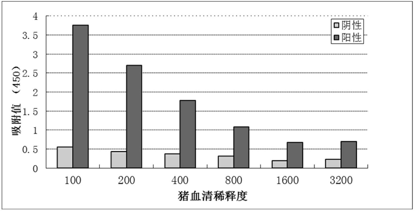
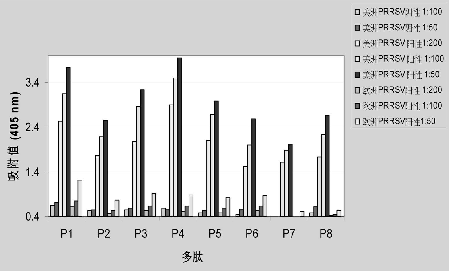
[0046] The selection and determination of the effective polypeptide segment is to predict the epitope of the porcine PRRS virus protein by general computer analysis software, determine the amino acid sequence of the natural antigen, and then search for the epitopes targeted by the peptide segment. ELISA was used to detect the reaction of different polypeptides with porcine PRRS antiserum, and finally the polypeptides with good reactogenicity were selected.

[0047] In this example, according to the protein sequence of the American / Chinese type PRRSV (PRRSV), 8 polypeptides with good reactogenicity were screened out, and the sequences are as follows:

[0048] P1: KKEKKKTKSVKSLPGNKPVPC (SEQ ID NO: 1);

[0049] P2: KKCQPVKDSWMSSRGFDE (SEQ ID NO: 2);

[0050] P3: CSAGTGGADLPTDLPPKK (SEQ ID NO: 3);

[0051] P4: KRCSEDDHDDLGFMVPK (SEQ ID NO: 4);

[0052] P5: CGFMVPPGLSSEGHLTKK (SEQ ID NO: 5);

[0053] P6: CLKSLVLGGRKAVKQGKK (SEQ ID NO: 6);

[...


PUM


Abstract

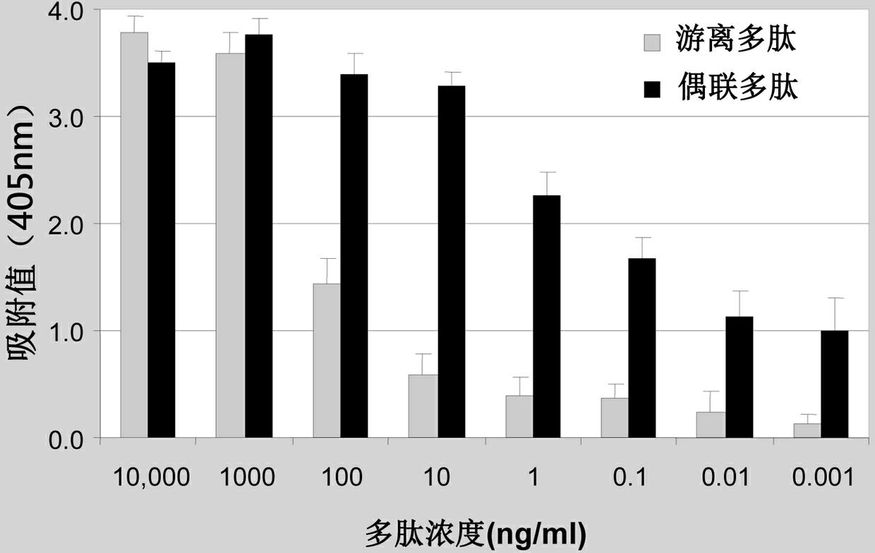
The invention discloses a virus infection detection method. The virus infection detection method comprises the following steps of: (1) coupling a viral reactogenicity polypeptide to a non-protein polymer to obtain a coupled polypeptide; and (2) adding the coupled polypeptide in a coating liquid, then adding a serum sample (to be detected) to perform the ELISA (enzyme-linked immunosorbent assay) detection, judging the virus and subtype infection thereof according to an immunoadsorption reaction. The detection method provided by the invention can obviously improve the detection sensitivity as well as obviously reduces the non-specific background readout, has high specificity, simplicity in operation, rapidness in diagnosis speed, and is economic and convenient in large scale detection; and the detection method is a good method easy for popularization and wide in application prospect. In the detection provided by the invention, the dosage of the coating antigen can be 10 ng / ml, and the less dosage is in favor of popularization and application.

Description

technical field [0001] The invention relates to a method for detecting virus infection, in particular to a method for detecting virus infection using a polypeptide as an antigen. Background technique [0002] At present, the diagnostic methods for viral diseases around the world mainly include virus isolation, molecular biology diagnosis (RT-PCR), immunoperoxidase cell monolayer assay (IPMA) based on serological tests, and neutralization test (SN). , indirect immunofluorescence test (ILFT), and enzyme-linked immunosorbent assay (ELISA) and so on. Compared with other methods, ELISA detection is easy to operate, easy to standardize, fast and sensitive, and is very suitable for large-scale epidemic investigation and disease surveillance. ELISA detection is divided into indirect ELISA and competition ELISA. In traditional ELISA detection, the amount of coated antigen is large, generally 5-10 μg / ml. Due to the large amount of antigen used, the detection cost is relatively high...

Claims


Application Information

Patent Timeline
Patent Type & Authority Applications(China)
IPC IPC(8): G01N33/569
Inventor 李其昌王贵平陈克宏
Owner GUANGDONG HAID ANIMAL HUSBANDRY & VETERINARY RES INST
Learn More