
Cell selection apparatus, and cell selection method using the same

A screening device and screening method technology, applied in the field of cell screening, can solve the problems of unable to screen cells, complicated separation procedures, etc.

Inactive Publication Date: 2011-05-18
TOSOH CORP
View PDF8 Cites 8 Cited by
  • Summary
  • Abstract
  • Description
  • Claims
  • Application Information

AI Technical Summary

Problems solved by technology

However, there is a problem that the process of separating target cells from captured cells becomes complicated
And in this method, since the cells are screened by whether the binding occurs through the antigen-antibody reaction, the cells cannot be screened based on the size of the binding force in the antigen-antibody reaction.

Method used


Image

  • Cell selection apparatus, and cell selection method using the same
  • Cell selection apparatus, and cell selection method using the same
  • Cell selection apparatus, and cell selection method using the same

Examples


Embodiment 1

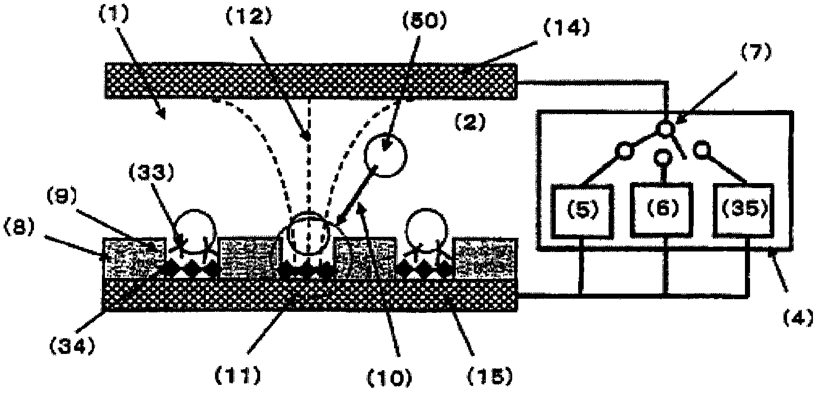
[0237] Figure 4 A schematic diagram of the cell screening device used in Example 1 is shown. Roughly divided, the cell screening device consists of a cell screening container (13) and a power supply (4). Cell screening containers such as Figure 4 The structure shown is as follows: a spacer (16) is arranged between an upper electrode (14) and a lower electrode (15), and an insulator (8) having a plurality of fine holes formed in an array is sandwiched between the spacer and the lower electrode . Also, as will be described later, micropores are formed in the insulator disposed on the lower electrode (15) by general photolithography and etching.

[0238] As the upper electrode and the lower electrode, electrodes formed of ITO (film thickness 150 nm) on a Pyrex (registered trademark) substrate with a length of 70 mm x a width of 40 mm x a thickness of 1 mm were used. A silicone rubber sheet having a shape of 20 mm in length by 20 mm in width and having a shape of 40 mm in le...

Embodiment 2

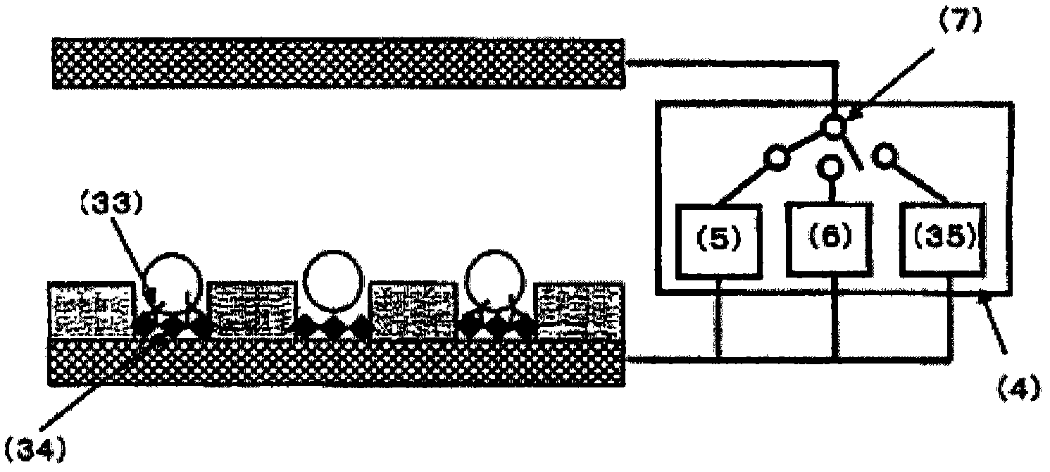
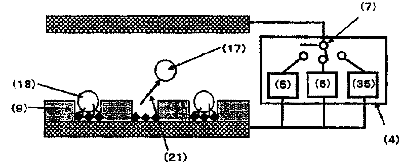
[0255] Antibody-immobilized polystyrene beads used in Example 1 to immobilize the anti-E2 antibody on the surface with the anti-E2-mouse antibody solution at each concentration of 0, 0.1, 0.5, and 2.0 μg / mL were introduced into the spacer. 600 μL of the suspension (bead concentration: about 1.65 x 10 6 cells / mL) were introduced into the cell screening area with a 1mL volume dispenser, and left to stand for about 5 minutes to allow the antibody-immobilized polystyrene beads to settle by gravity, and then left to stand at room temperature for about 40 minutes to allow the beads to enter the micropores. The antibody-immobilized polystyrene beads in the method are in contact with the bottom surface of the micropore, so that the antibody on the surface of the antibody-immobilized polystyrene bead reacts with the antigen immobilized on the bottom surface of the micropore. Next, an AC voltage of 2.5 Vpp and a frequency of 3 MHz was applied between the electrodes by the second AC powe...

Embodiment 3

[0261] Using the same cell screening apparatus as in Example 1, spleen cells having specific antibodies present on their surfaces were screened. As the cells, non-immunized mouse spleen cells A and mouse spleen cells B immunized with the E2 antigen were used. In addition, in the spleen cells B of mice immunized with the E2 antigen, there were cells in which the E2 antibody was present on the cell surface (hereinafter referred to as antibody-containing cells). This Example 3 is an example of identifying the antibody present in the cell and the antigen immobilized on the bottom surface of the micropore by the magnitude of the alternating voltage of the second alternating current power supply for determining the magnitude of the negative dielectrophoretic force. The binding force of the antigen-antibody reaction, thereby screening the cells for the presence of antibodies.

[0262] First, mouse spleen cells A and mouse spleen cells B were respectively suspended in 300 mM mannitol...


PUM

PropertyMeasurementUnit
diameteraaaaaaaaaa
diameteraaaaaaaaaa

Abstract

Disclosed are a cell selection apparatus and a cell selection method for efficiently selecting a large quantity of cells capable of producing a specific substance in one step, within a short period and in a simple manner, based on the strength of binding force between the specific substance on the surface of each of the cells and a recognizing molecule capable of recognizing the substance. The cell selection apparatus comprises: a pair of electrodes; a cell selection vessel which is made of a sheet-like insulating material having multiple micropores and has a recognizing molecule arranged on the bottom of each of the micropores, wherein the recognizing molecule can bind to the specific substance; and an electric power supply. In the cell selection apparatus, the electric power supply has an electric power supply for immobilizing cells and an electric power supply for collecting cells. The cell selection method uses the cell selection apparatus. In the cell selection method, cells are introduced into a cell selection zone, the cells are immobilized in the micropores, and the binding reaction between the specific substance on the surface of each of the cells and the recognizing molecule is carried out. Subsequently, a cell in which the specific substance on the surface of the cell is not bound to the recognition molecule or a cell in which the specific substance is weakly bound to the recognition molecule is removed from the micropores, or alternatively, a cell in which the specific substance on the surface of the cell is strongly bound to the recognition molecule is left in the micropores.

Description

technical field [0001] The present invention relates to a cell screening device for efficiently performing cell screening, and a cell screening method using the same. [0002] This application claims the priority of Japanese Patent Application No. 2008-105396 for which it applied in Japan on April 15, 2008, and uses the content here. Background technique [0003] In recent years, the use of antibodies produced in vivo has attracted attention as diagnostic reagents for diseases and raw materials for pharmaceuticals. An antibody is a type of protein that specifically binds to a foreign substance such as a virus when it invades a living body. Antibodies bind to foreign substances such as specific viruses to kill or detoxify the viruses, and have the function of protecting organisms from invasion by viruses and the like. Here, a foreign substance such as a virus to which an antibody specifically binds is generally referred to as an antigen. In addition, antibodies in the livi...

Claims


Application Information

Patent Timeline
Patent Type & Authority Applications(China)
IPC IPC(8): C12M1/00C12M1/42C12Q1/04G01N33/53
CPCG01N33/5438G01N33/569
Inventor 山中麻帆丸山高广二见达
Owner TOSOH CORP
Learn More