
Method for establishing lactation model of cow mammary gland epithelial cells

A technology for mammary epithelial cells and cells, which is applied in the field of biological cytology, can solve the problems of immature in vitro culture technology of dairy cow mammary epithelial cells, and achieve the effects of low culture cost, low culture condition requirements and high survival rate.

Inactive Publication Date: 2010-02-24
NORTHEAST AGRICULTURAL UNIVERSITY
View PDF0 Cites 7 Cited by
  • Summary
  • Abstract
  • Description
  • Claims
  • Application Information

AI Technical Summary

Problems solved by technology

In vitro culture technology for dairy cow mammary epithelial cells has been immature

Method used


Image

  • Method for establishing lactation model of cow mammary gland epithelial cells
  • Method for establishing lactation model of cow mammary gland epithelial cells
  • Method for establishing lactation model of cow mammary gland epithelial cells

Examples


Embodiment 1

[0024] Embodiment 1: a kind of method of the present invention establishes cow mammary gland epithelial cell lactation model, the experimental method step is as follows:

[0025] Step 1: Primary culture and identification of dairy cow mammary gland epithelial cells

[0026] Disinfect the middle part of the breast, cut out a small amount of breast tissue according to conventional surgery, immediately wash the tissue with D-Hanks solution and peel off the fat tissue and connective tissue as much as possible, and cut the breast acinar tissue into 1mm 3 Inoculate small pieces of collagen in the cell culture flask pre-coated with collagen at a distance of 0.5 cm. After covering the entire bottle, culture it upside down at 37°C 2 In the incubator for about 3-4 hours, until the tissue block is firmly attached to the collagen, gently add the growth medium to the culture, so as to cover the tissue block, then place it in the incubator to continue the culture, and replace the culture ev...

Embodiment 2

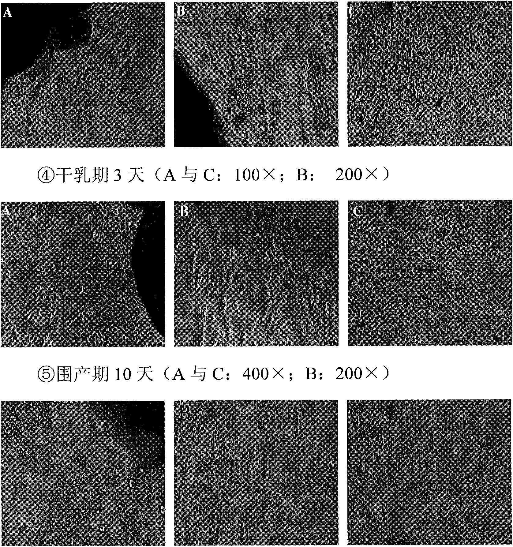
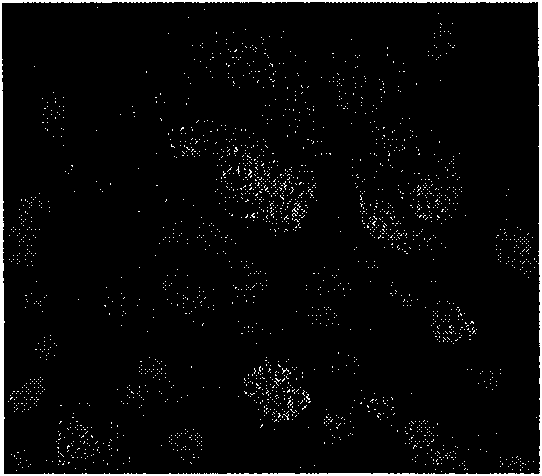
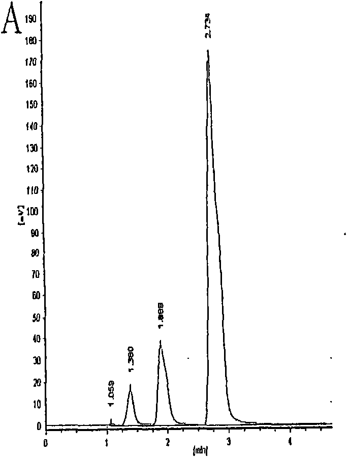
[0033] Example 2: Combining Figure 1-Figure 6 , the experiment done by the present invention:

[0034] Experimental material: healthy Holstein cow mammary gland tissue. The breast tissues sampled were in puberty for 2 months, lactation period for 7 days, lactation period for 280 days, dry period for 3 days, and perinatal period for 10 days. Experimental reagents: rat tail collagen, Hangzhou Shengyou; high-quality fetal bovine serum, DF12 medium, Gibco; cytokeratin 18 antibody, Acris, Germany; PI dye, insulin, Sigma; chromatography grade methanol, Tianjin Guangfu; lactose standard Products, Academy of Military Medical Sciences; β-casein standard, Sigma. Experimental equipment: ZHJH-1112 vertical flow ultra-clean bench, Shanghai Zhicheng Analytical Instrument Manufacturing Co., Ltd.; CO-150 CO 2 Incubator, Japan; SZ-93 automatic double pure water distiller, Shanghai Yarong Biochemical Instrument Factory; LC-10AT high performance liquid chromatography, Shimadzu, Japan, SPD-10...


PUM


Abstract

The invention provides a method for establishing a lactation model of cow mammary gland epithelial cells for researching the milk protein gene expression regulation mechanism and providing favorable conditions for testing a mammary gland expression vector. The testing method comprises the following steps: carrying out primary culture and identification of the cow mammary gland epithelial cells; determining the secretion situation of lactose by using the HPLC method; determining the secretion situation of beta-casein protein by using the HPLC method; and carrying out cryopreservation and recovery on the cells. The method adopts the tissue block method to culturing the cow mammary gland epithelial cells in different periods, compared with other primary culture methods of the mammary gland epithelial cells, such as enzyme digestion and the like, the method does not affect the activity of the mammary gland epithelial cells, the culture cost is very low, and the operation is very simple. The method realizes the continuation of normal culture of a cow mammary gland epithelial cell system, thereby property and effectively preserving a large number of precious cow mammary gland tissue materials. The method provides the important testing materials and a technical platform for researching the lactation mechanism of the cow mammary gland during the different development periods.

Description

(1) Technical field [0001] The invention relates to biological cytology, in particular to a method for establishing a lactation model of cow mammary gland epithelial cells. (2) Background technology [0002] Mammary gland epithelial cells are the basic structural and functional units of lactation. By studying the expression of lactation-related genes in them, it is possible to reveal how mammary gland-specific expression genes are activated, how important functional genes for lactation initiate and maintain lactation, and regulate milk components. Theoretical basis. At the same time, it is helpful to establish a mammary gland bioregulation technology that can regulate the expression level of important functional genes in dairy cow lactation and significantly increase milk production. [0003] Primary culture refers to the period from the time when tissue cells are removed from the body to the first passage, which is a characteristic and inevitable growth stage. Cells or ti...

Claims


Application Information

Patent Timeline
IPC IPC(8): C12N5/06G01N30/02A01N1/02
Inventor 佟慧丽高学军李庆章林叶
Owner NORTHEAST AGRICULTURAL UNIVERSITY
Learn More