Method for diagnosing cancer through detection of deglycosylation of glycoprotein
a glycoprotein and glycoprotein technology, applied in the direction of peptide/protein ingredients, peptide sources, instruments, etc., to achieve the effect of short total analysis time, high specificity and sensitivity, and efficient us
- Summary
- Abstract
- Description
- Claims
- Application Information
AI Technical Summary
Benefits of technology
Problems solved by technology
Method used
Image
Examples
example 1
of De-Glycosylated / Non-Glycosylated Peptides Using a Standard Glyco Proteins
[0078]The following experiments was performed using a standard protein to confirm the possibility of discovering or developing markers based on the quantification of glycosylated peptides and de-glycosylated peptides using MRM technology and its use in diagnostics.
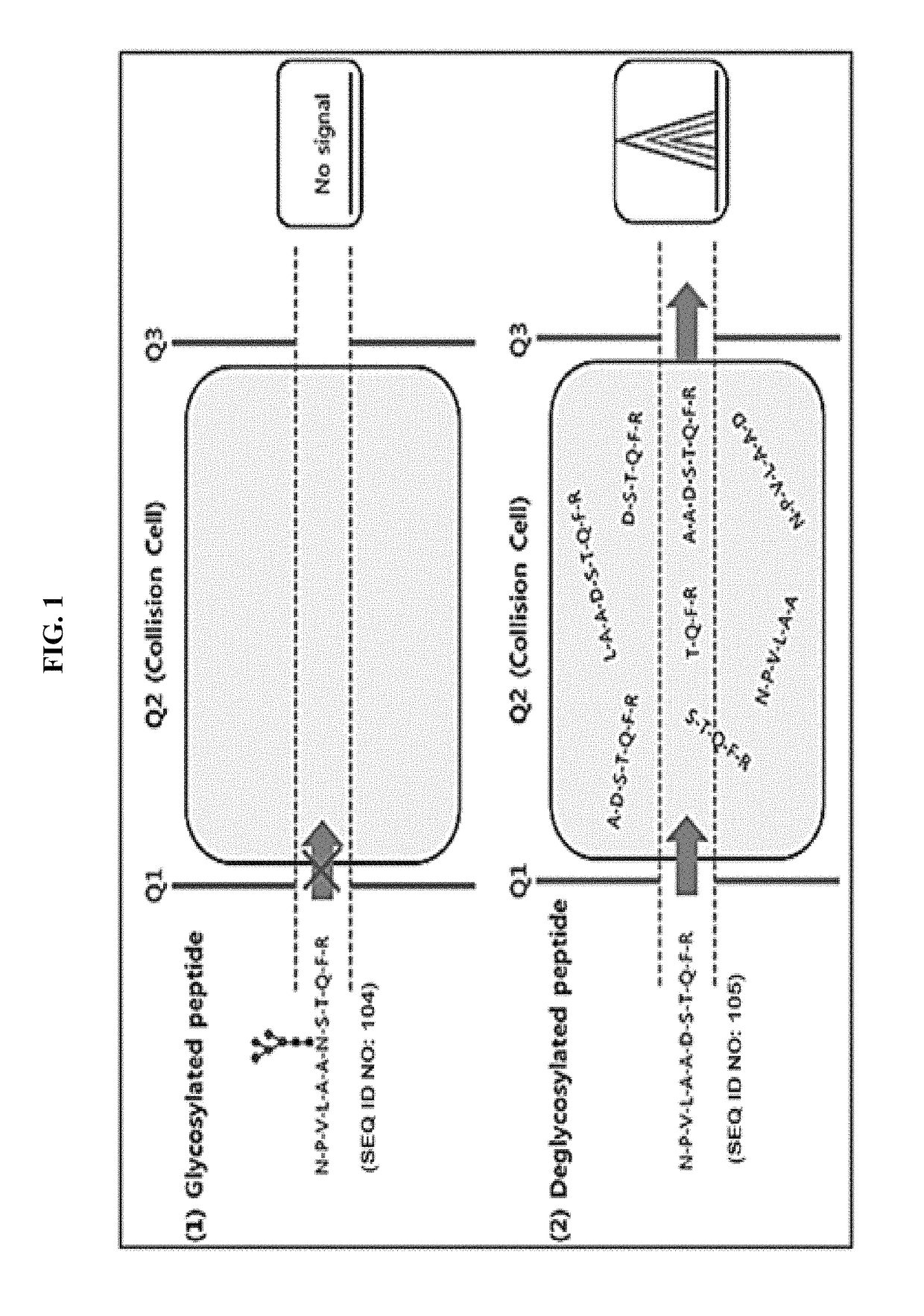
[0079]As shown in FIG. 1, MRM is a technology to quantify a relative or an absolute amount of proteins in biological fluids using Triple quadrupole as a mass spectrometry. MRM includes a first mass filter Quadruple 1 (Q1) filtering only the peptides with a specific m / z (mass / charge), Quadruple 2 (a collision cell) in which the peptides from Q1 are fragmented by electric energy and Quadruple 3 (Q3) which transmits only particular fragmented peptide ions. Then the ions transmitted through Q3 are shown as a peak of chromatogram at the detector. The area of the peak is calculated for the absolute or relative quantification of peptides. In case of glyco...
example 1-1
Standard Glycoproteins
[0080]Among the commercially available proteins which are purified and lyophilized, a protein in which both the glycosylated peptide having NxS / T motif and the non-glycosylated peptide without the motif are selected as predictable transitions in Skyline has been used as a standard.
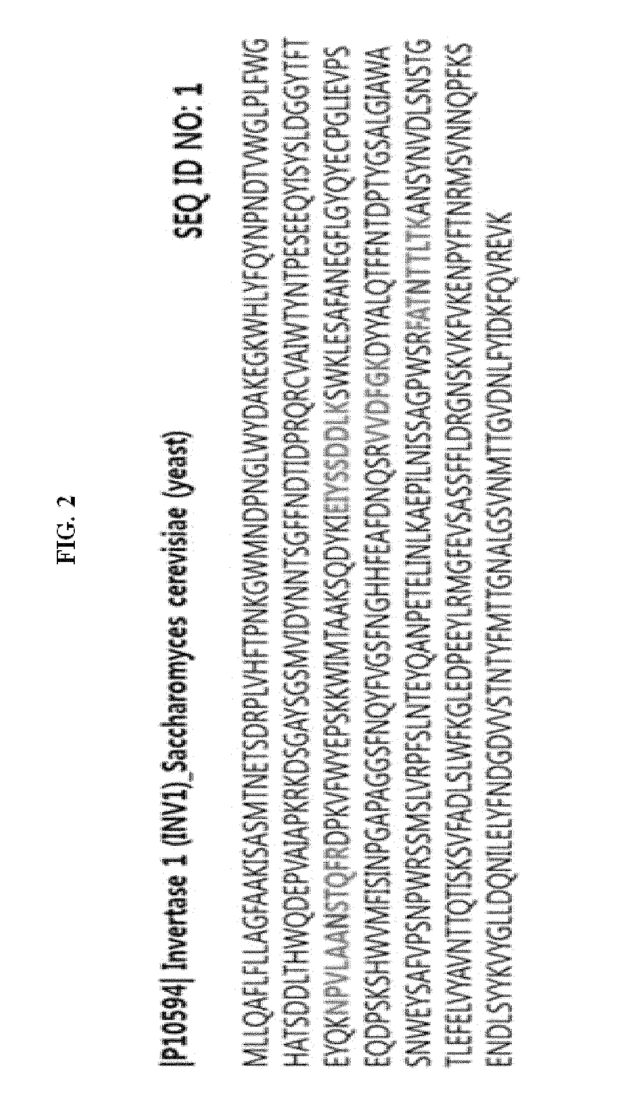
[0081]In the present example, Invertase-1protein was used as a standard and the sequence is as shown in FIG. 2 in which the green indicates the sequence used in the analysis. The sequence of the standard glycosylated peptides 1 and 2 employed are NPVLAANSTQFR (SEQ ID NO: 104) and FATNTTLTK (SEQ ID NO: 106), respectively. When the peptides are de-glycosylated, Asn residues are converted to Asp resulting in the peptide sequence of NPVLAADSTQFR (SEQ ID NO: 105) and FATDTTLTK (SEQ ID NO: 107), respectively. The standard non-glycosylated peptides 1 and 2 employed are IEIYSSDDLK (SEQ ID NO: 108) and VVDFGK (SEQ ID NO: 109), respectively.
example 1-2
Selection of the Theoretical Transition (Q1 / Q3) of the Standard Protein
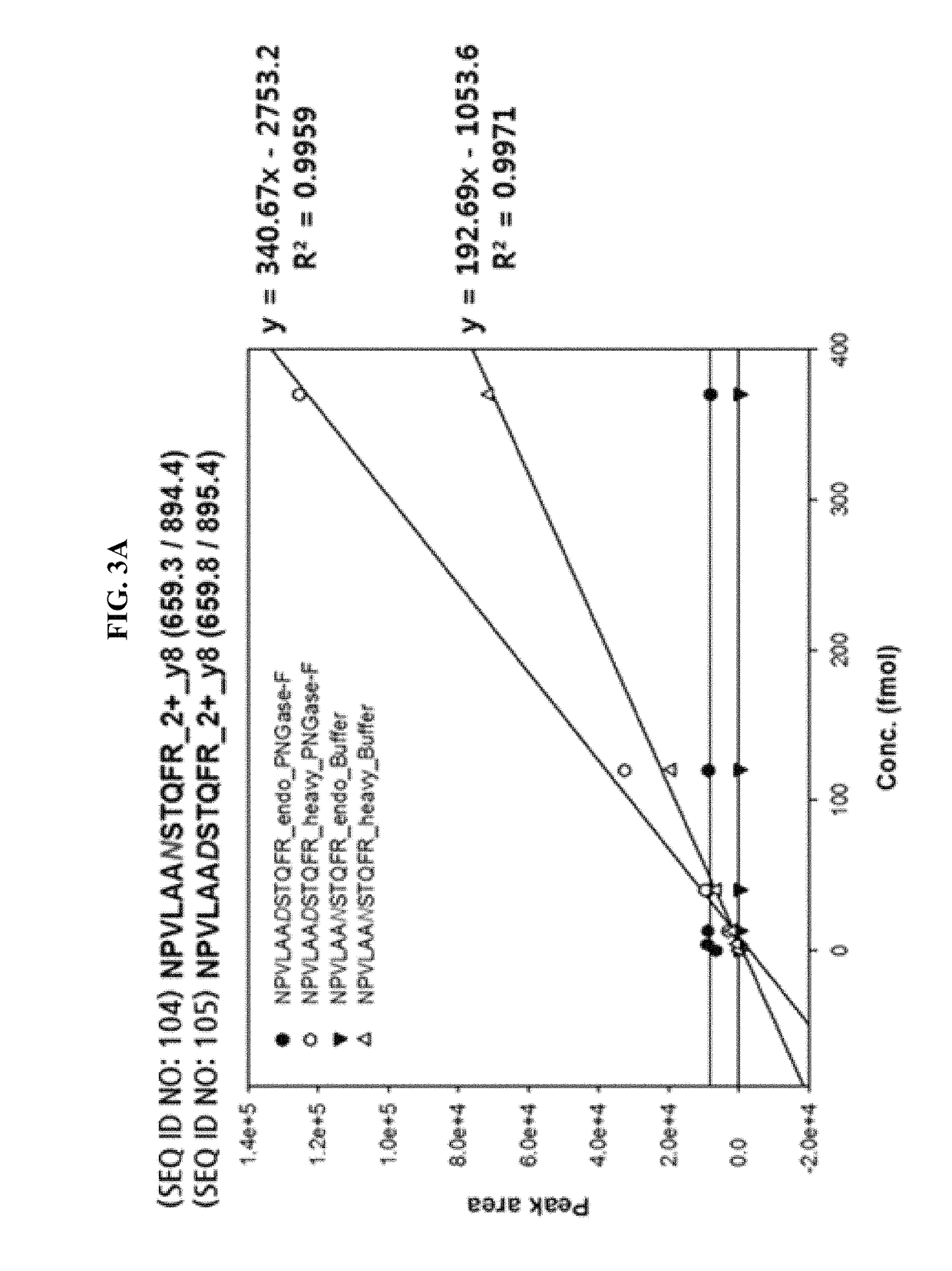
[0082]The native form of the sequence of the standard protein and the conversion form thereof in which N is changed to D at NxS / T motif were imported into Skyline (https: / / brendanx-uw1.gs.washington.edu / labkey / project / home / software / Skyline / begin.view) program to select a theoretical transition value. At the same time, synthetic peptides with the same sequence except that 12C and 14N atoms in Arg (R) and Lys (K) residues at the C-terminal region were heavy labelled with 13C and 15N were used to confirm that the peptides detected are actually from the peptides of interest to be detected.
[0083]That is, the heavy labelled peptide and the endogenous peptide share the same sequence and have the identical hydrophobicity. Thus they can be detected on LC-column (C18) since they are eluted at the same retention time.
[0084]As a result of the selection, Q1 difference of 0.49 Da and Q3 difference of 0.98 Da between the native...
PUM
| Property | Measurement | Unit |
|---|---|---|
| flow rate | aaaaa | aaaaa |
| temperature | aaaaa | aaaaa |
| temperature | aaaaa | aaaaa |
Abstract
Description
Claims
Application Information
 - R&D
- Intellectual Property
- Life Sciences
- Materials
- Tech Scout
- Unparalleled Data Quality
- Higher Quality Content
- 60% Fewer Hallucinations
Browse by: Latest US Patents, China's latest patents, Technical Efficacy Thesaurus, Application Domain, Technology Topic, Popular Technical Reports.
© 2025 PatSnap. All rights reserved.Legal|Privacy policy|Modern Slavery Act Transparency Statement|Sitemap|About US| Contact US: help@patsnap.com


