
Devices and methods for endovascular access and therapy

a technology for endovascular access and therapy, applied in the direction of catheters, applications, guide wires, etc., to achieve the effect of shortening the response tim

Active Publication Date: 2016-09-13
TRAUMATEK SOLUTIONS
View PDF67 Cites 23 Cited by
  • Summary
  • Abstract
  • Description
  • Claims
  • Application Information

AI Technical Summary

Benefits of technology

The invention provides devices and methods for quickly and safely accessing blood vessels to stop a bleed and stabilize a patient until definitive treatment can be provided. The cannulation device has a cannula with side openings and a distal tip, and may also include rollers or an expandable sheath. The device can also include a support with magnetic field detectors to help locate the catheter tip. The vessel cannulation device has a lumen and may also include a pressure sensor, an automatic guidewire release mechanism, a wheel mechanism for pushing a guidewire, a needle, and an expandable sheath. Overall, this invention helps improve trauma teams' ability to manage blood vessel access and save lives.

Problems solved by technology

Although extremely common, the ways to establish vascular access remain very basic and are often inadequate.
This is especially unfortunate in elective settings, as it is those older and sicker individuals who usually have more “difficult blood vessels”, and must frequently endure additional suffering caused by painful repeated attempts at blood vessel cannulation, even when performed by experienced personnel.
However the emergency setting also increases the obstacles to successful blood vessel cannulation.
A venous cut down may be performed using simple tools by an experienced physician, but this too takes time and requires certain expertise, making it impractical in many cases.
Massive hemorrhage remains the major cause of death in the battlefield.
The Seldinger technique, although useful, suffers from several drawbacks.
First, it requires significant experience in order to be successfully performed, especially when circumstances are suboptimal such as in emergency and trauma situations.
Second, there are several points during the procedure which may lead to its failure.
Additionally, this might cause blood to leak around the vessel causing an internal hematoma, which might compress the vessel and make repeat cannulation more difficult.
Worse yet, unintended movement of the needle might place it within one of the arterial walls, and attempted insertion of the guide wire can then damage the arterial wall, possibly leading to large hematomas or other complications.
Exit of the guide-wire from the artery at this stage will cause the sheath to be placed into tissues instead of into the artery, which besides tissue damage usually causes the guide-wire to bend, necessitating its replacement.
Additional drawbacks of the Seldinger technique are related to the use of a long guide-wire, which carries with it an increased risk of contamination of its proximal end, as well as a danger of splashing blood at the physician.
Also, during the time between needle entry into the vessel and until the guide-wire is inserted into it, either profuse bleeding or entry of air into the circulation might occur, depending on whether pressure within the vessel is higher or lower than ambient pressure.
Since the “over-the-needle” technique is not adequate for placing large bore catheters or sheaths, the Seldinger technique is used in these cases, which as mentioned, include most endovascular interventions.
The WAND mainly addresses safety issues such as needle-stick injury and air embolism but the technique is still rather complicated and requires significant training.
Such solutions are cumbersome and expensive and are not appropriate for direct over the needle vascular access.
Another drawback of existing sheaths related to their having a fixed diameter, is that the arterial puncture site remains dilated to the maximum size for the whole duration of the procedure.
First, in patients with hemorrhagic shock, aortic cross clamping redistributes the patient's limited blood volume to the myocardium and brain.
This may in part be because EDT is an aggressive procedure, and its invasive nature and associated morbidity limit its therapeutic potential.
Possible risks and complications of EDT include:technical complications which may include but are not limited to: unintentional injury to the heart, coronary arteries, aorta, phrenic nerves, esophagus, and lungs, avulsion of aortic branches to components of the mediastinum;compromised respiratory function;increased risk for hypothermia;recurrent chest bleeding;infection of the pericardium, pleural spaces, sternum, and chest wall;post-pericardiotomy syndrome; andhigh risk of personnel exposure to HIV or hepatitis.
When using traditional equipment, EAO requires experience with endovascular techniques as well as specialized equipment such as fluoroscopy, both of which are usually unavailable in the resource limited battlefield environment or even in the ED.

Method used


Image

  • Devices and methods for endovascular access and therapy
  • Devices and methods for endovascular access and therapy
  • Devices and methods for endovascular access and therapy

Examples


embodiment 10

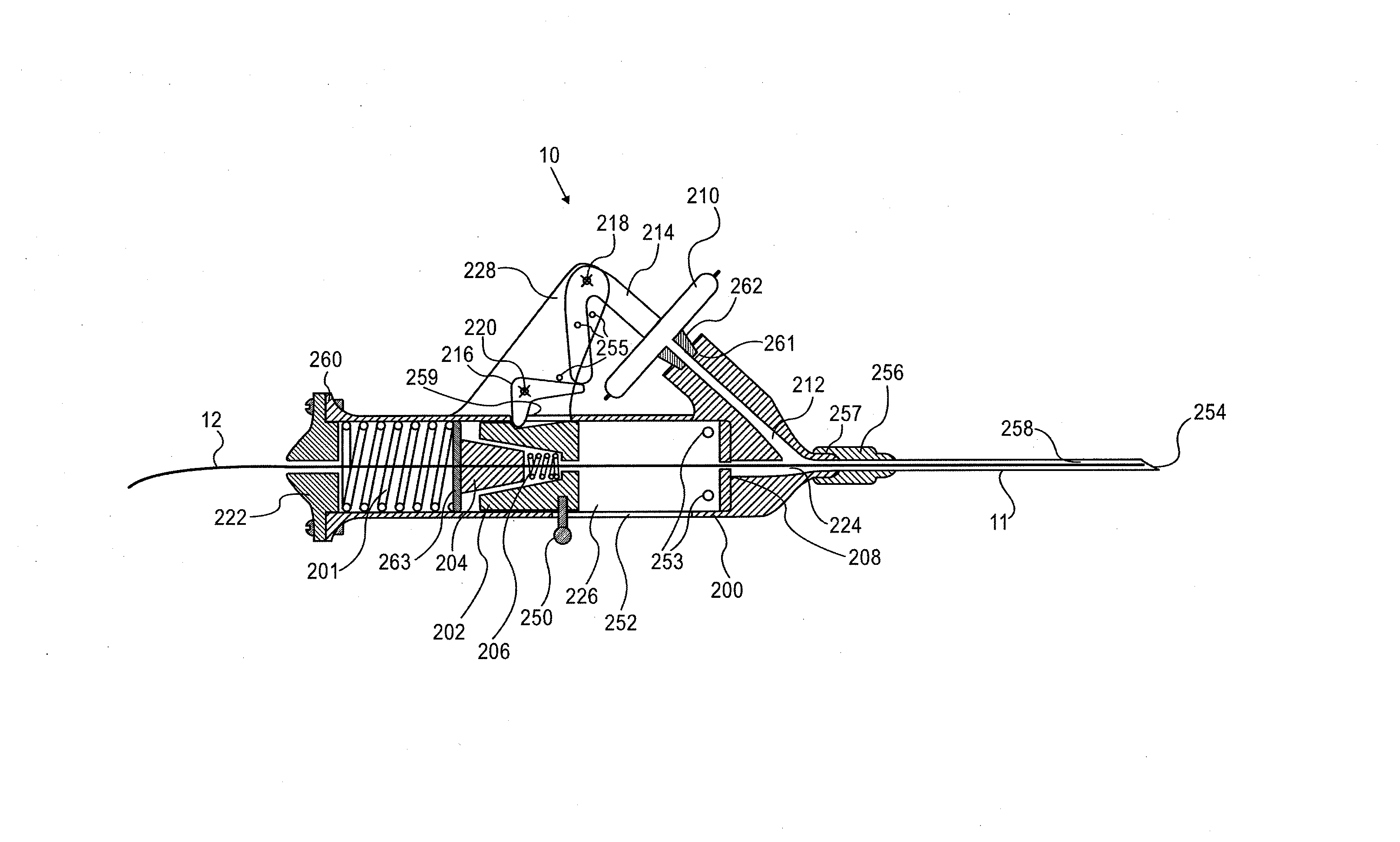
[0133]A preferred embodiment of the vessel cannulation device is a spring-loaded device, which automatically inserts a guidewire into the vessel upon penetration of the vessel by a needle. An embodiment 10 of such a device is shown in FIG. 3A-C. More particularly, FIG. 3A is a longitudinal section of an embodiment of vessel cannulation device 10 in its loaded / armed state, including a body 200, with needle 11 having needle tip 254, needle lumen 258 and needle hub 256 is attached to its front end at needle adapter 257. Body 200 includes needle adapter 257, anterior lumen 224, channel 212 ending in body to diaphragm interface 261, posterior lumen 226, air release holes 253, window 252, aperture 259, and bracket 228. Backplate 222 is connected to proximal end 260 of body 200. Diaphragm 210 connects to body 200 at body to diaphragm interface 261 through diaphragm adaptor 262. Diaphragm 210 is in fluid communication with adapter 262, body to diaphragm interface 261, channel 212, needle ad...

embodiment 2500

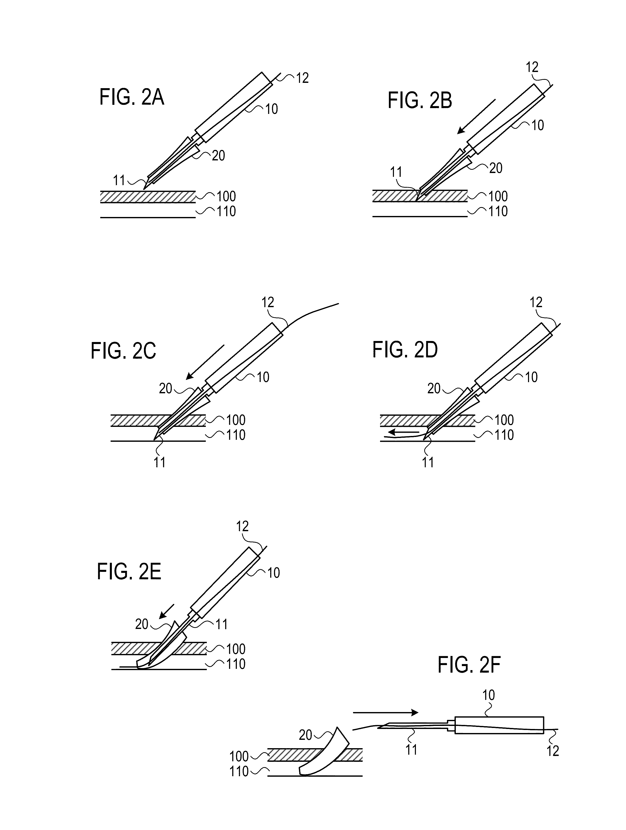
[0185]Embodiment 2500 of vessel cannulation device shown in FIGS. 42A-B utilizes a similar mechanism to that previously described for vessel cannulation device 10, in order to automatically advance a sheath over the needle into the blood vessel. Device 2500 automatically advances a sheath over the needle a large distance, inserting the sheath into the vessel.

[0186]Typically, in embodiment 2500, sheath 2512 will be automatically advanced between about 20 mm-100 mm, preferably about 30 mm-60 mm.

[0187]More particularly, FIG. 42A is a longitudinal section of an embodiment of vessel cannulation device 2500 in its loaded / armed state, including a body 200, with needle 11 having needle tip 254, needle lumen 258 and needle hub 256 is attached to its front end at needle adapter 257. Body 200 includes needle adapter 257, anterior lumen 224, channel 212 ending in body to diaphragm interface 261, posterior lumen 226, air release holes 253, window 252, aperture 259, and bracket 228. Backplate 222...


PUM


Abstract

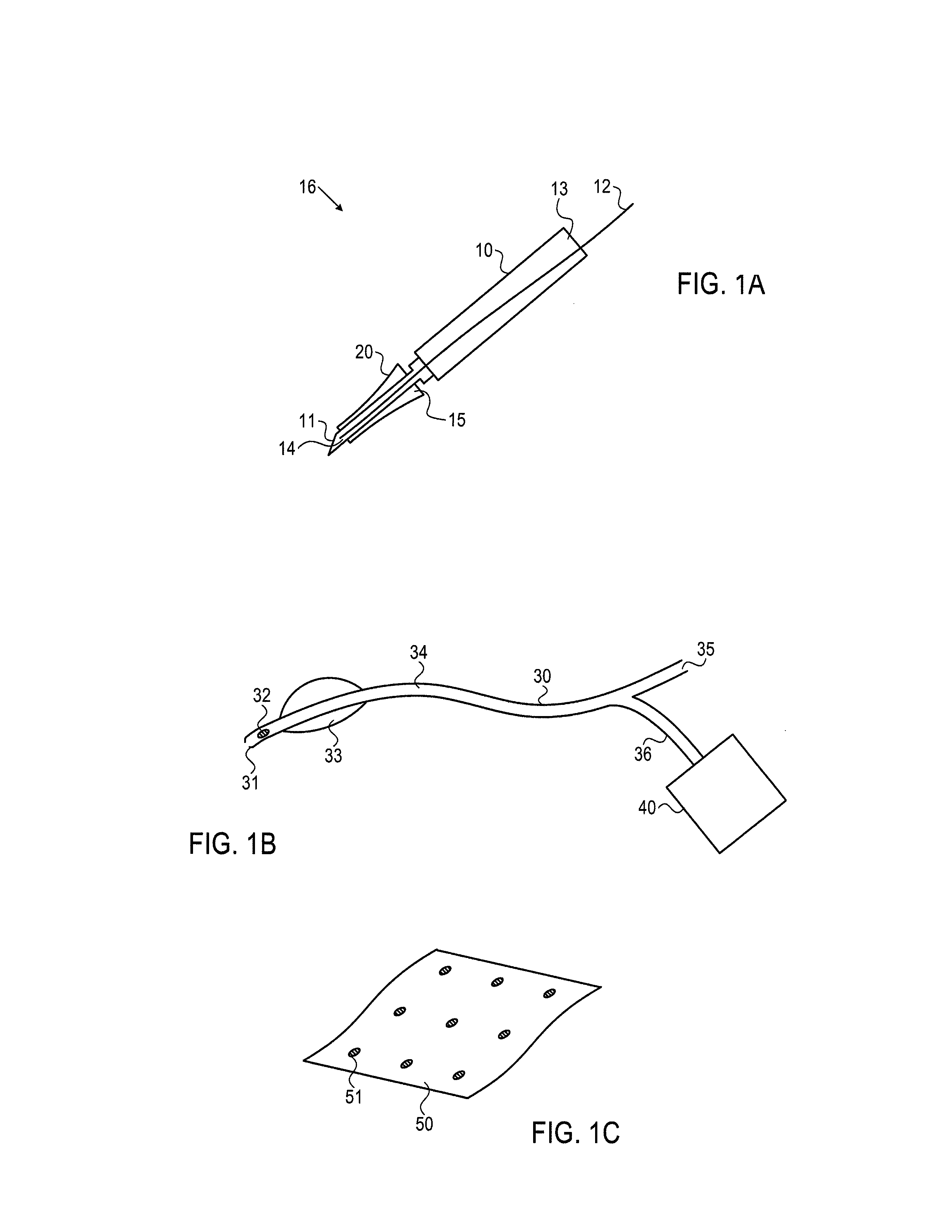
The present invention provides for devices and methods for providing endovascular therapy, including facilitating establishment of vascular access, placement of endovascular sheaths, catheter tip localization, and administration of vascular occlusion. The inventions includes a vessel cannulation device, an expandable sheath, an occlusion catheter, and a localizer each of which may be provided separately or used as part of a system.

Description

CROSS-REFERENCE TO RELATED APPLICATIONS[0001]This application claims priority to U.S. Provisional Application 61 / 568,111 (filed on Dec. 7, 2011) and U.S. Provisional Application No. 61 / 612,334 (Mar. 18, 2012), the disclosures of which are incorporated by reference in their entireties.FIELD OF THE INVENTION[0002]The invention relates to devices and methods for providing endovascular therapy, including facilitating establishment of vascular access, placement of endovascular sheaths, catheter tip localization, and administration of vascular occlusion.BACKGROUND OF THE INVENTIONI. Clinical Need[0003]General:[0004]Vascular access is a crucial element of medical therapy in a vast majority of clinical settings and procedures. This is true in both elective and in emergent situations. In a specific type of emergency, hemorrhagic shock, there may further be a need to perform aortic occlusion. Both these clinical needs, vascular access and aortic occlusion, are the subject of the current inven...

Claims


Application Information

Patent Timeline
Patent Type & Authority Patents(United States)
IPC IPC(8): A61B17/12A61B17/34A61M25/06A61M25/01A61B17/00A61M25/10A61M25/09A61M25/00
CPCA61B17/12109A61B17/12031A61B17/3403A61M25/0105A61M25/065A61B17/1214A61B17/12136A61B17/12168A61B2017/00022A61B2034/2072A61M25/0127A61M25/09041A61M2025/0024A61M2025/1052A61B2090/064A61B46/10A61B2034/2051A61B2090/3954A61M25/0023
Inventor AVNERI, ITZHAKAVNERI, BEN-AMIAVNERI, SHAHARAVNERI, LIOR
Owner TRAUMATEK SOLUTIONS
Learn More