
Expression and assembly of human group C rotavirus-like particles and uses thereof

a technology of rotaviruses and particles, applied in the field of group c rotaviruses, can solve the problems of low detection rate of gpc rv, insensitivity to gpc rv, and inability to use adequate diagnostic tools

Active Publication Date: 2015-10-27
UNITED STATES OF AMERICA
View PDF8 Cites 3 Cited by
  • Summary
  • Abstract
  • Description
  • Claims
  • Application Information

AI Technical Summary

Benefits of technology

The present invention provides isolated recombinant human rotavirus group C virus-like particles that include a human rotavirus group C VP6 protein, a human rotavirus group C VP7 protein, and optionally a human rotavirus group C VP2 protein. These virus-like particles are free of other human rotavirus group C proteins such as VP1, VP3, VP4, NSP1, NSP2, NSP3, NSP4, NSP5, NSP6, and NSP7. The invention also provides processes for detecting a human rotavirus group C antibody, delivering a cargo moiety to a cell, and generating an immunological response in a human. The isolated polypeptide and nucleic acid molecule of the invention include the amino acid sequence of the human rotavirus group C VP6 protein, the amino acid sequence of the human rotavirus group C VP7 protein, and optionally the amino acid sequence of the human rotavirus group C VP2 protein.

Problems solved by technology

One possible cause of low GpC RV detection is the unavailability of adequate diagnostic tools.
While PCR is a frequently employed technique, it is often insensitive for diagnosis of GpC RV due to the instability of its capsid proteins and the degradation of its RNA genome.
It is also not an accessible technique to many clinical laboratories that are involved in diagnostics of samples from patients with gastroenteritis.
However their specificity and sensitivity to human GpC RV is questionable.
Progress in the development of a sensitive and specific EIA for human GpC RV has been stunted by its fastidious growth in cell culture.
To the best of our knowledge, these reagents have not been utilized for viral detection in human specimens and their specificity to GpC RV remains questionable.
However these diagnostic tools may not be sensitive or specific enough for human strains.
Thus, sensitive and specific detection methods and reagents for human group C RV are not readily available.
Consequently, the burden of GpC RV disease has not been clearly established.

Method used


Image

  • Expression and assembly of human group C rotavirus-like particles and uses thereof
  • Expression and assembly of human group C rotavirus-like particles and uses thereof
  • Expression and assembly of human group C rotavirus-like particles and uses thereof

Examples


example 1

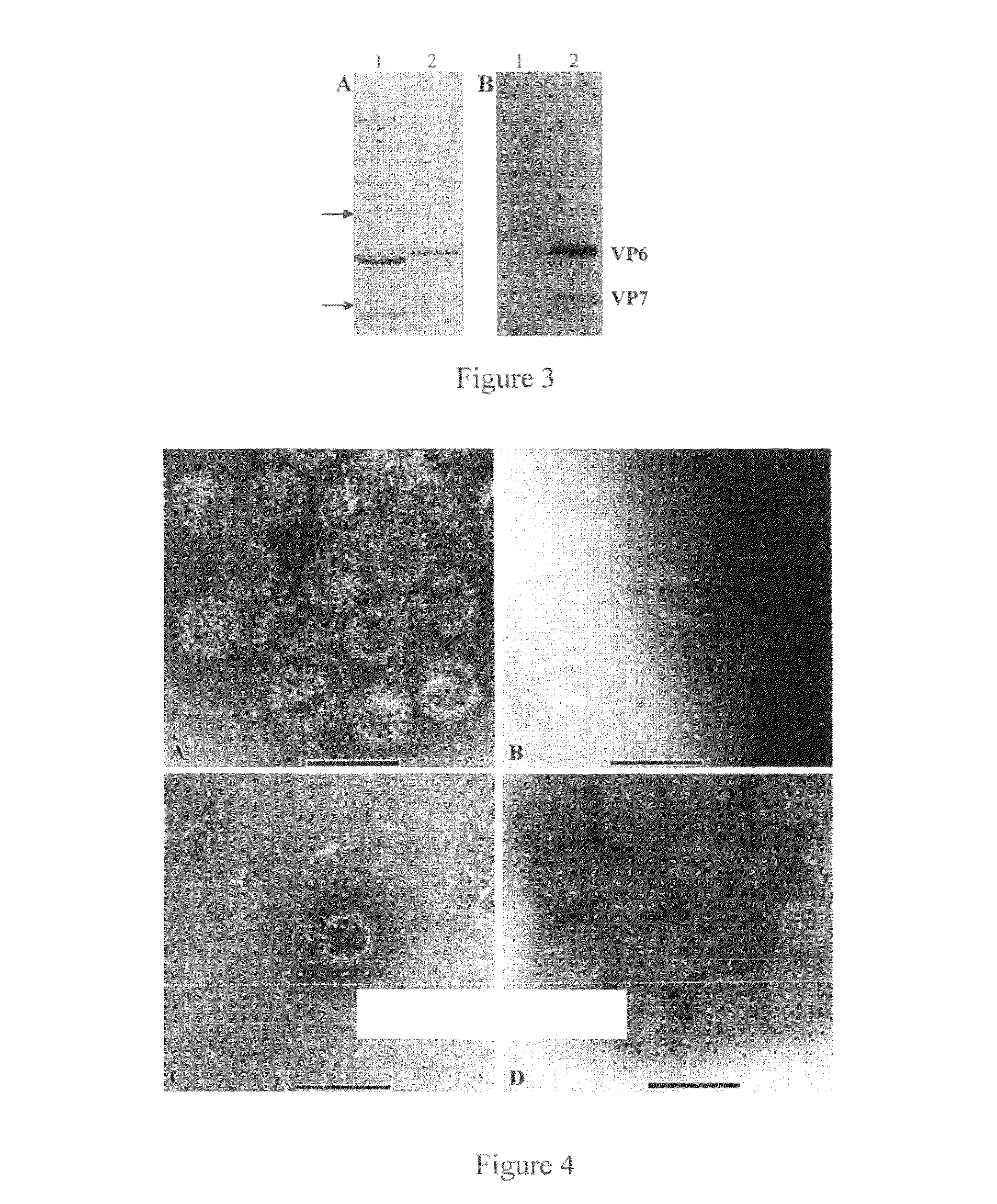
[0204]Cloning and construction of baculovirus recombinants. Segment 5, encoding VP6, from human group C RV strain S-1 was amplified by RT-PCR using BMJ44 (5′-AGC-CAC-ATA-GTT-CAC-ATT-TC-3′) (SEQ ID NO: 14) and BMJ141 (5′-ATC-TCA-TTC-ACA-ATG-GAT-G-3′) (SEQ ID NO: 15) (28). Segment 8, encoding VP7, from strain S-1 was amplified by RT-PCR using primers BMJ13 (5′-AGC-CAC-ATG-ATC-TTG-TTT-3′) (SEQ ID NO: 20) and BMJ14 (5′-GGC-ATT-TAA-AAA-AGA-AGA-3′) (SEQ ID NO: 21) (13, 28). Segment 2, encoding VP2, from strain ASP88 was amplified by RT-PCR using BMJ197 (5′-TCG-AGG-ACA-AAT-CGT-CCA-AG-3′) (SEQ ID NO: 22) and BMJ180 (5′-AGC-CAC-AGA-GTT-TGA-GGT-C-3′) (SEQ ID NO: 23). Cloning and construction of recombinant baculovirus expressing S-1 VP7 was previously described (14). DNA fragments of segment 2 and 5 were cloned into vector pVL1393 and transfections were performed with the Bac-N-Blue transfection kit (Gibco, Grand Island, N.Y.). Baculovirus constructs were amplified in Spodoptera frugiperda 9 ...

example 2

[0211]Cells and superinfections. Sf9 or High Five (Hi5) insect cells were grown and maintained in EX-CELL 420 or 405 media (Sigma, Lenexa, Kans.) or HyQ SFX-INSECT media in shaker flasks at 27° C. Sf9 and Hi5 cells were subcultured every 3 or 4 days at a concentration of 1×10⁶ cells / ml and 5×10⁵ cells / ml, respectively. Stationary superinfections were performed by seeding Sf9 cells in HyQ or EX-CELL 420 and Hi5 cells in HyQ or EX-CELL 405 into a T150 flask at a concentration of 3×10⁵ cells / ml. Baculovirus constructs (rVP2, rVP6 and rVP7) were added at a multiplicity of infection (MOI) of 1 each. Infections were carried out without proteinase inhibitors and infected cultures were harvested at day 5. Large scale VLP production was performed in suspension culture by seeding Sf9 cells in EX-CELL 420 into fernbach flasks at a concentration of 1×10⁶ cells / ml. Baculovirus recombinants were added one day later at an MOI of 1.4 each and harvested on day 4.

example 3

[0212]Western blot. Sodium dodecyl sulfate-polyacrylamide gel electrophoresis (SDS-PAGE) was carried out with 12% separating and 5% stacking gels using the Laemmli discontinuous buffer system (16). Samples were heated at 97° C. for 5 min with 10% β-mecaptoethanol prior to loading and then electrophoresed. Proteins were transferred to a PVDF membrane in transfer buffer (25 mM Tris, 192 mM glycine, 10% methanol). After blocking with 10% (for unpurified GpC RV proteins) blotto in PBS-T for 1-2 hrs at room temperature or 15% (for purified GpC RV proteins) blotto in PBS-T overnight at 4° C., membranes were incubated with porcine hyperimmune serum (1:2,000) to Cowden in 5% blotto in PBS-T overnight at 4° C. or rabbit hyperimmune serum (1:20,000) to human GpC VLPs in 10% blotto in PBS-T for 1 h. Membranes were washed in PBS-T, incubated with horseradish peroxidase (HRP) goat anti-pig (1:142,000) (KPL, Gaithersburg, Md.) in 5% blotto or HRP-goat anti rabbit (1:20,000) (Pierce, Rockford, Ill...


PUM

PropertyMeasurementUnit
temperatureaaaaaaaaaa
pHaaaaaaaaaa
pHaaaaaaaaaa

Abstract

Group C rotaviruses are a cause of acute gastroenteritis in children and adults that is distinct from group A RV. However, human group C rotaviruses cannot be grown in culture, resulting in a lack of tools for detection and treatment of GrpC RV disease. Consequently, the burden of GpC RV disease has not been clearly established. Isolated recombinant human rotavirus group C virus-like particles are provided according to embodiments of the present invention along with methods of their production and use in, inter alia, detection of Grp C RV infection, diagnostic assays and immunogenic compositions.

Description

REFERENCE TO RELATED APPLICATIONS[0001]This application claims priority from U.S. Provisional Patent Application Ser. No. 61 / 130,615 filed May 29, 2008 and U.S. Provisional Patent Application Ser. No. 61 / 111,425 filed Nov. 5, 2008, the entire content of both are incorporated herein by reference.GOVERNMENT SPONSORSHIP[0002]This invention was made by the Centers for Disease Control and Prevention, an agency of the United States Government.FIELD OF THE INVENTION[0003]The present invention relates to group C rotaviruses (GpC RV) and rotavirus-like particles, methods of producing immunogenic rotavirus-like particles, immunogenic compositions inclusive of rotavirus-like particles and methods for eliciting an immune response using compositions inclusive of rotavirus-like particles, as well as producing a diagnostic for rotavirus infection.BACKGROUND OF THE INVENTION[0004]Rotaviruses are a diverse set of pathogens classified into groups A through G based on the distinct characteristics of t...

Claims


Application Information

Patent Timeline
Patent Type & Authority Patents(United States)
IPC IPC(8): A61K39/12C12N7/02C12N7/00C07K14/005A61K39/00
CPCC07K14/005A61K39/12A61K2039/5258C12N2710/14143C12N2720/12322C12N2720/12323C12N2720/12334A61P31/14A61P37/04
Inventor JIANG, BAOMING
Owner UNITED STATES OF AMERICA
Learn More