
Volumetric deformable registration method for thoracic 4-D computed tomography images and method of determining regional lung function

a volumetric deformation and computed tomography technology, applied in image enhancement, image analysis, instruments, etc., can solve the problems of reduced compliance, thicker and stiffer tissue, and associated mechanical changes within the lung itsel

Active Publication Date: 2015-07-07
UNIV OF LOUISVILLE RES FOUND INC
View PDF1 Cites 43 Cited by
  • Summary
  • Abstract
  • Description
  • Claims
  • Application Information

AI Technical Summary

Benefits of technology

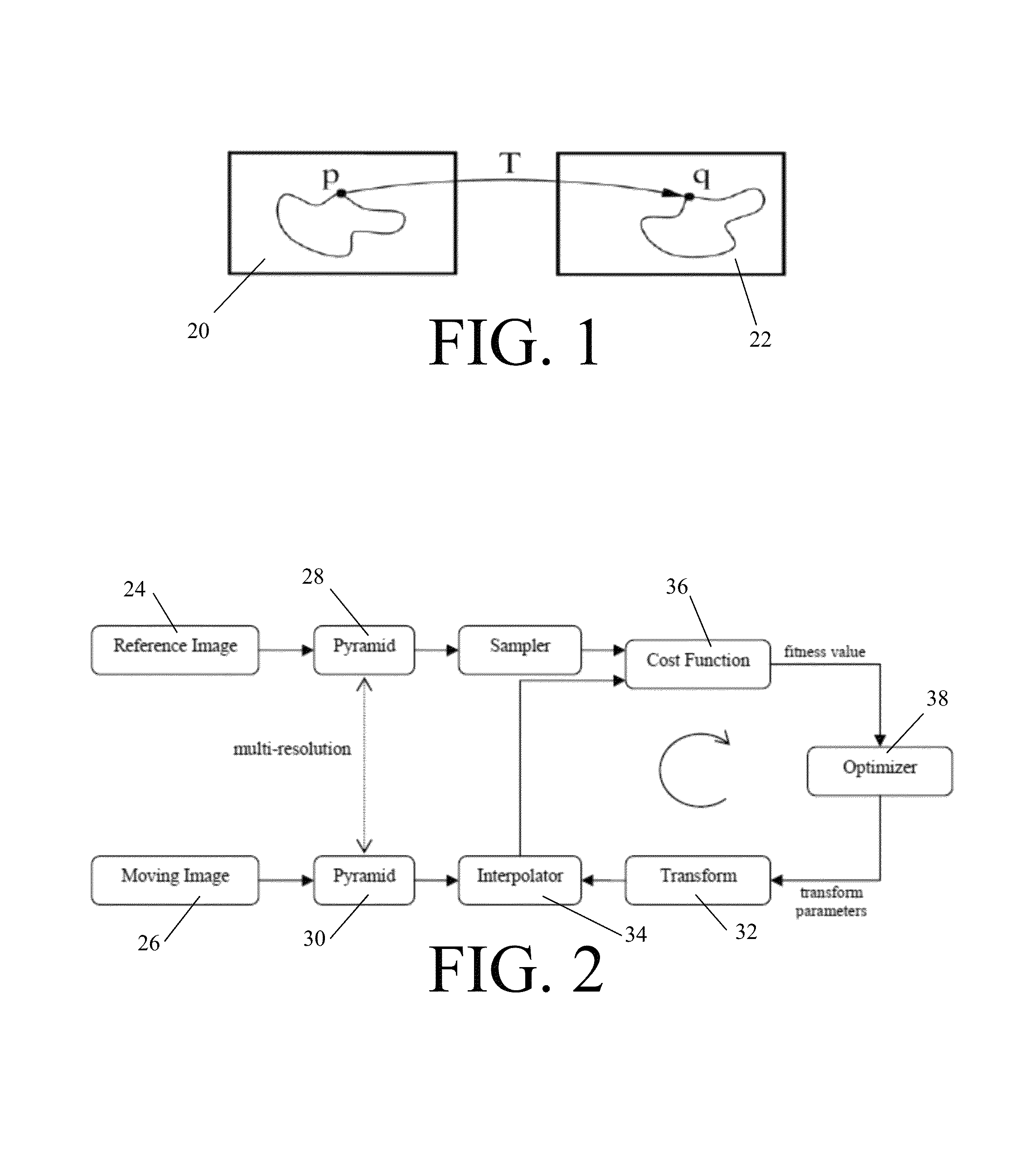
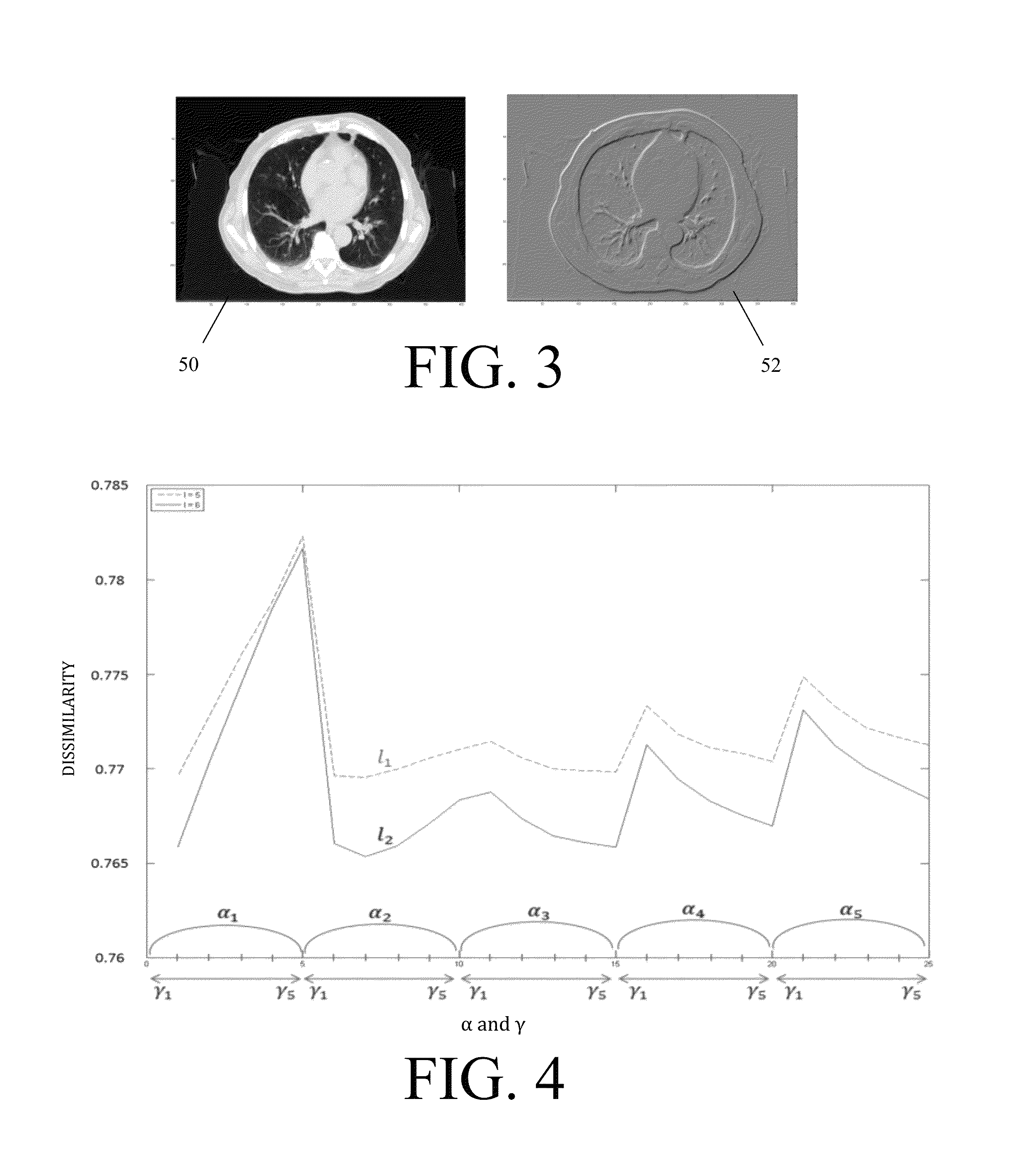
The present invention provides objective methods for registering and analyzing volumetric deformation in thoracic 4-D computed tomography (CT) images. These methods involve solving an energy function to transform the images into a smooth motion field, while enforcing constraints on the images to ensure smoothness and consistency. The invention also provides methods for determining the regional mechanics of the lung by analyzing the strain patterns in the image data. These methods can help improve the accuracy and reliability of lung function analysis and diagnosis.

Problems solved by technology

The mechanical properties of lung parenchymal tissue are both elastic and dissipative, as well as being highly nonlinear.
As another example, idiopathic pulmonary fibrosis (IPF), a classic interstitial lung disease, causes inflammation and fibrosis of tissue in the lungs and, over time, the disease makes the tissue thicker and stiffer (reduced compliance) and subsequently leads to associated mechanical changes within the lung itself.
Despite advances in CT imaging, however, respiratory motion often degrades anatomic position reproducibility during imaging.
In turn, that degradation then necessitates larger margins to be drawn during certain therapy planning, such as during radiotherapy planning where smaller margins can cause errors during radiation delivery.
In all of these types of radiation therapy, however, large margins around a tumor must typically be drawn as the imaging often prevents precise anatomical mapping of the tumor and healthy tissue.
Furthermore, the image acquisition process often renders the resulting thoracic CT images prone to noise, blurring, and image artifacts.
Moreover, motion reproducibility assumption with respect to the breathing index and insufficient number of projections per breathing phase for volumetric 3-D reconstruction continue to be frequent limitations of 4-D CT.

Method used


Image

  • Volumetric deformable registration method for thoracic 4-D computed tomography images and method of determining regional lung function
  • Volumetric deformable registration method for thoracic 4-D computed tomography images and method of determining regional lung function
  • Volumetric deformable registration method for thoracic 4-D computed tomography images and method of determining regional lung function

Examples


example 1

Multi-Scale Optical Flow Method Incorporating Mass Conservation for Deformable Motion Estimation

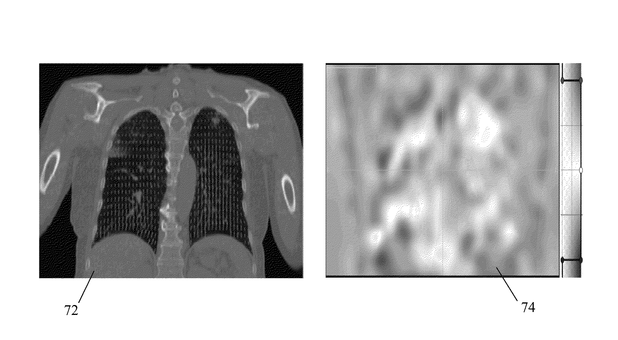
[0072]For both qualitative and quantitative evaluation of the exemplary method described herein, the publicly available POPI-model (J. Vandemeulebroucke, D. Sarrut, and P. Clarysse, “The POPI-model, a point-validated pixel-based breathing thorax model,” in International Conference on the Use of Computers in Radiation Therapy (ICCR), Toronto, Canada, 2007) and DIR-lab (R. Castillo, E. Castillo, R. Guerra et al., “A framework for evaluation of deformable image registration spatial accuracy using large landmark point sets,” Physics in medicine and biology, vol. 54, no. 7, pp. 1849-1870, 2009) data of lung deformation has been used. In the POPI-model dataset, 41 homologous landmarks were defined by experts in each of the ten respiratory phases of a single individual that make up this 4-D CT dataset with voxel dimensions 0.97×0.97×2 mm and made up of 512×512×141 voxels. For the DIR-lab data, 7...

example 2

Method of Determining Regional Lung Function

[0091]Seven patients with non-small cell lung cancer (NSCLC) who were scheduled to receive thoracic radiotherapy were enrolled in a study and imaging was performed prior to the initiation of treatment. 4-D CT data were collected with 1.17×1.17×3 mm voxel dimension using a Philips Brilliance Big Bore CT scanner and the Varian Real-time Position Management (RPM) system (Varian Medical Systems, Palo Alto, Calif.) to record patient respiratory traces in the Department of Radiation Oncology at University of Louisville. An audiovisual feedback device was utilized to ensure a reproducible and consistent respiratory cycle waveform to ensure fidelity of the 4-D CT data. For each patient, 4-D CT images of the entire thorax and upper abdomen were obtained.

[0092]Each patient also received tomographic SPECT ventilation / perfusion lung imaging on a Philips ADAC Sky-light Dual head gamma camera. For SPECT ventilation, Tc-99m DTPA was aerosolized and inhal...


PUM


Abstract

A method of deformable image registration for thoracic 4-D computed tomography (CT) images includes: receiving, by a processing device, a set of thoracic 4-D CT images; and iteratively solving, by the processing device, an energy function applied to subsequent images of the set of thoracic 4-D CT images to transform the subsequent images into respective optical flow fields between the subsequent images, the energy function enforcing the following constraints on the subsequent images: intensity constancy; mass conservation; gradient constancy; and spatio-temporal smoothness. A method of determining regional lung function includes: receiving, by a processing device, a set of thoracic 4-D CT images of a lung; transforming, by the processing device, the set of 4-D CT images of the lung into respective spatial voxel-wise deformation maps; and transforming, by the processing device, the spatial voxel-wise deformation maps into respective spatial voxel-wise strain maps of the lung indicating regional mechanics of the lung.

Description

RELATED APPLICATIONS[0001]This application claims priority to U.S. Provisional Patent Application No. 61 / 618,081, filed Mar. 30, 2012, the entire disclosure of which is incorporated herein by reference.TECHNICAL FIELD[0002]The presently-disclosed subject matter relates to a volumetric deformable registration method for thoracic 4-D computed tomography (CT) images and to a method of determining regional lung function. In particular, the presently-disclosed subject matter relates to a volumetric deformable registration method and a method of determining regional lung function whereby a set of thoracic 4-D CT images of lungs are transformed into optical flow fields and / or deformation maps.BACKGROUND[0003]Lungs are essential organs for respiration, facilitating gas exchange between the human body and the atmosphere by delivering oxygen from the air into the venous blood and extracting carbon dioxide from the blood at the same air-blood interface. Moreover, lungs play other critical role...

Claims


Application Information

Patent Timeline
IPC IPC(8): G06T7/00
CPCG06T7/0012G06T7/0024G06T2207/10076G06T2207/30061G06T2207/10081G06T7/30G06T7/38
Inventor NEGAHDAR, MOHAMMADRREZAAMINI, AMIR A.DUNLAP, NEALWOO, SHIAO
Owner UNIV OF LOUISVILLE RES FOUND INC
Learn More