Tomosynthesis imaging

a technology of tomosynthesis and imaging, applied in the field of xray-based imaging systems and methods, can solve the problems of increasing patient discomfort, physical movement, and increasing the time of image acquisition, and achieve the effect of reducing the fluence rate of x-rays and high x-ray fluence ra

Active Publication Date: 2014-08-26
UNIV OF MASSACHUSETTS
View PDF19 Cites 10 Cited by
  • Summary
  • Abstract
  • Description
  • Claims
  • Application Information

AI Technical Summary

Benefits of technology

[0035]In another embodiment, the x-ray source configured to provide a first x-ray beam having a high x-ray fluence rate is a high power source.

Problems solved by technology

Although this is technically attainable, the physical movement of the tube is the source of many problems in tomosynthesis.
A moving x-ray tube prolongs the exposure time and the duration of physical compression of the breast that in turn increases patient discomfort.
Moreover, the resulting longer image acquisition time is more likely to contribute to blurring of the images due to patient motion and physical movement of the x-ray tube.
However, such systems lack a central high power x-ray tube or other type of high-power x-ray source.
The fixed multi-spot x-ray sources are significantly underpowered and currently they are not capable of delivering the high x-ray fluence needed for mammography in an acceptable time frame to minimize patient motion.
Therefore, such systems will be limited to tomosynthesis use only and not mammography.
However, a mammographic or radiographic unit that can only operate in the tomosynthesis mode is too limited and it will not be desirable in most medical practices, because most breast imaging centers would prefer a system that can perform both tests.
This rapid firing and mechanical repositioning creates many problems that lead to a less than optimal tomosynthesis image acquisition.
The tube current and x-ray output can be hard to control without elaborate and expensive electronic controls.
Moreover, the x-ray filament is susceptible to the mechanical vibrations of the movement of the tube and this can have a negative effect on the spatial resolution of images.
A mechanical “stop-fire-and-go” approach is very problematic because of the mechanical instabilities due to acceleration and deceleration of the mechanical assembly of the x-ray source.
Continuous mechanical movement is generally preferred but it also prone to vibrations that can affect the image quality.
In addition, during each firing that is of finite duration, the x-ray tube is in continuous motion resulting in blurring that degrades image quality.
It typically takes between four to ten seconds for a complete acquisition and this increases the chances for a slight movement of the breast or other part of the body that will degrade the spatial resolution and diagnostic quality of the images.
This motion problem can be minimized by applying additional compression on the breast using the pneumatic compression mechanism and plate but this is highly undesirable because of increased pain or discomfort.
In chest and abdominal radiography this problem is even more serious because shorter exposures are required, typically in the order of milliseconds in chest imaging.
A number of problems in attempting to implement a method and system that provides both radiographic and tomographic images have been observed.

Method used


Image

  • Tomosynthesis imaging
  • Tomosynthesis imaging
  • Tomosynthesis imaging

Examples


Embodiment Construction

Combined Digital Mammography with High Speed Tomosynthesis

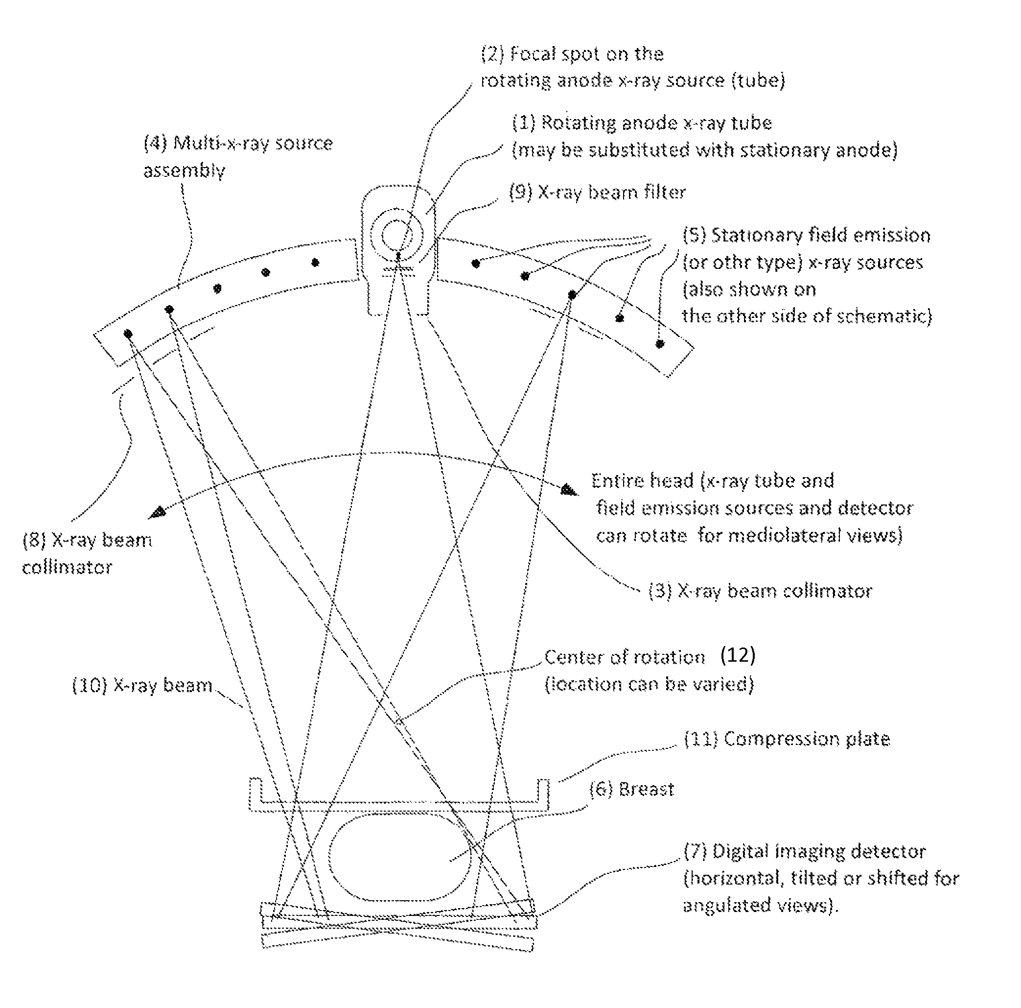
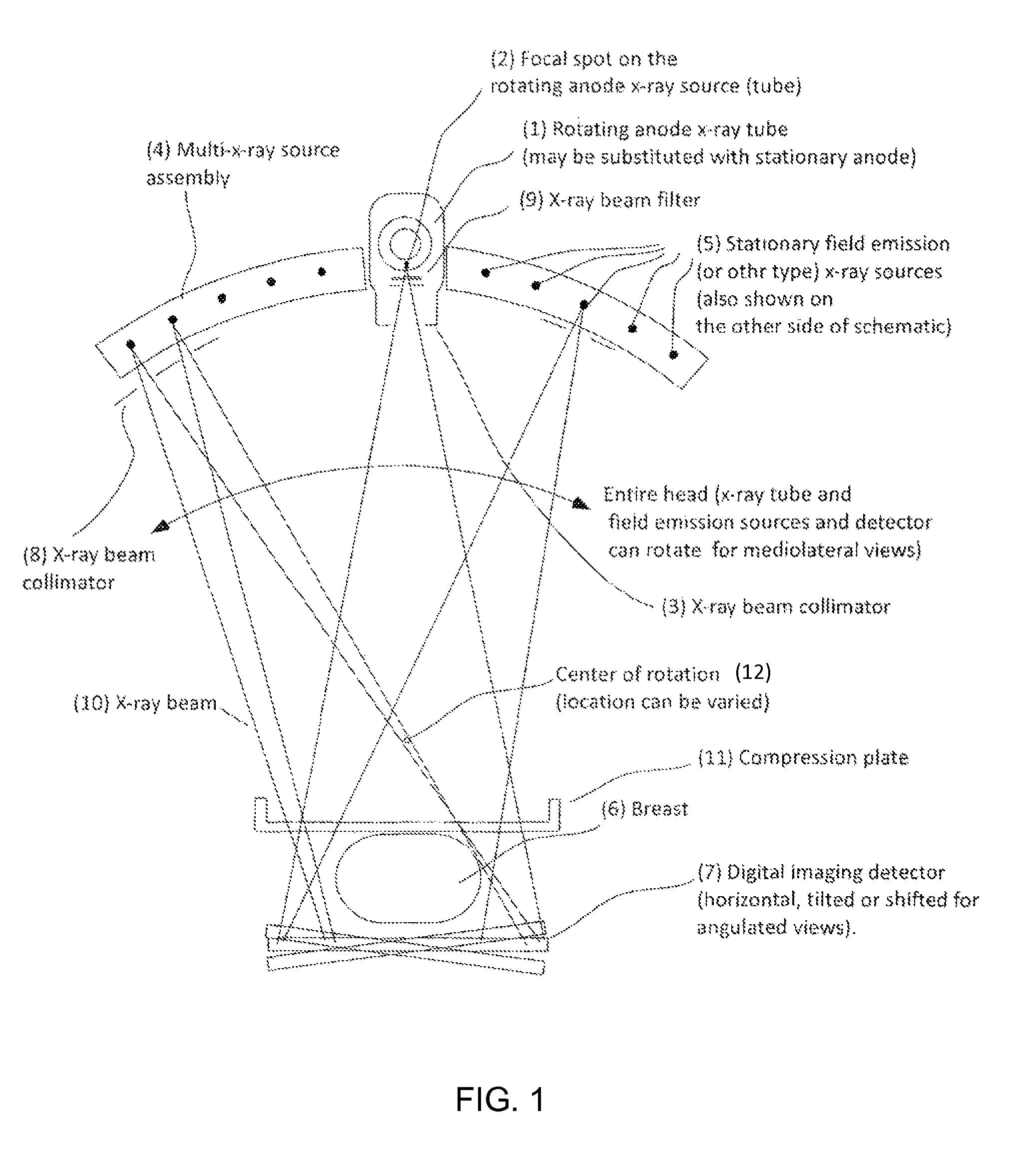
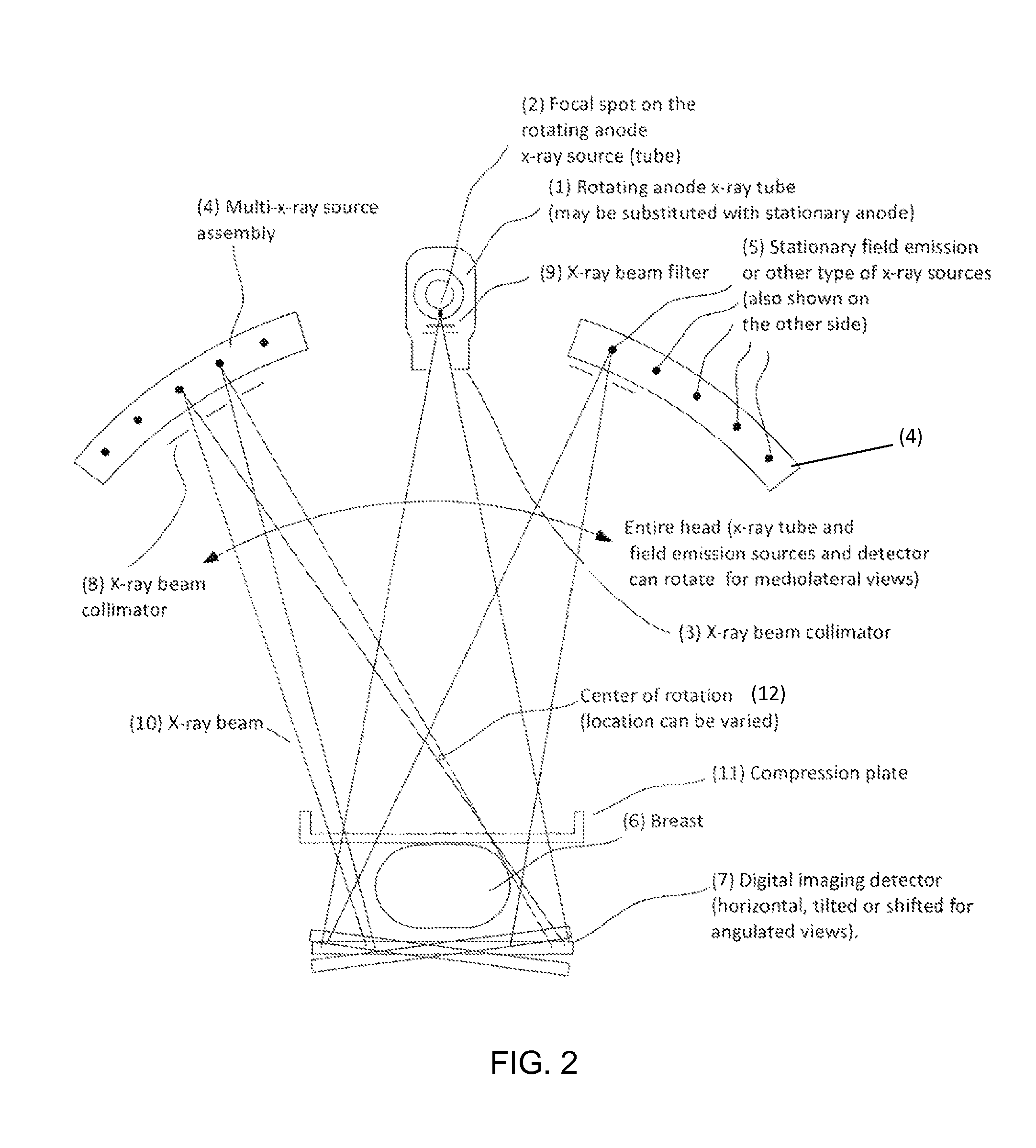
[0058]We describe a method for tomosynthesis imaging that uses a number of x-ray sources positioned on each side (or only on one side if desired) of the conventional central x-ray tube. This preferred embodiment enables fast tomographic image acquisition for tomosynthesis and for stereoscopic x-ray imaging. Image acquisition parameters (kVp, target / filter, mAs), either individually or in combination, can be varied with projection angle during acquisition of the (n+1) projections. We refer to such variation of operating parameters as angle adaptive beam modulation (AABM).

[0059]In order to overcome the limitations of prior art systems, it is desirable and advantageous to have a series of spatially fixed x-ray sources in an arc path that can activate sequentially or in any desired order to cover the desired angle of exposure in the shortest possible time.

[0060]We describe a system that is expected to be fully capable of operatin...


PUM


Abstract

Systems and methods for providing radiographic, stereoscopic and tomographic images of an object of interest. Examples of objects of interest are body parts of living beings, such as the human breast and the human chest. The apparatus includes a high-fluence rate x-ray source and a plurality of satellite x-ray sources operating at lower fluence rate than the high-fluence rate source. A controller controls the operation and locations of the sources, and the operation of a detector. The method provides procedures in which the operation of the high-fluence source and the satellite sources are individually controlled as to location and orientation relative to the object of interest. In some operations, one satellite source may be operating while another satellite source may be repositioning. By proper control, a reduced x-ray dose and reduced operating time can be attained, thereby improving image quality, patient care, and patient experience.

Description

CROSS-REFERENCE TO RELATED APPLICATIONS[0001]This application claims priority to and the benefit of U.S. provisional patent application Ser. No. 61 / 437,749 filed Jan. 31, 2011 which application is incorporated herein by reference in its entirety.STATEMENT REGARDING FEDERALLY FUNDED RESEARCH OR DEVELOPMENT[0002]This invention was made with government support under Grant Nos. CA134128, CA128906 and EB004015 awarded by the National Institutes of Health. The Government has certain rights in the invention.FIELD OF THE INVENTION[0003]The invention relates to x-ray-based imaging systems and methods in general and particularly to an imaging system and method that provides x-ray images.BACKGROUND OF THE INVENTION[0004]Digital radiography provides a two-dimensional (2-D) image of a three-dimensional (3-D) object resulting in superposition of structures. Stereoscopic imaging is a technique wherein at least two 2-D x-ray projection images, referred to as an image pair, separated by an angle not...

Claims


Application Information

Patent Timeline
Patent Type & Authority Patents(United States)
IPC IPC(8): A61B6/00A61B6/02
CPCA61B6/022A61B6/502A61B6/4007A61B6/025A61B6/4233A61B6/4291A61B6/5282A61B6/0414
Inventor VEDANTHAM, SRINIVASANKARELLAS, ANDREW
Owner UNIV OF MASSACHUSETTS
Why Eureka
Try Eureka