
Methods of use for an immunoassay detecting fragment Ba

a technology of immunoassay and fragment, applied in the field of new techniques for prenatal diagnosis, can solve the problems of adverse pregnancy outcomes, uncontrolled complement activation, and inflammation of the placenta, and achieve the effects of reducing the risk of infection, and reducing the effect of immunoassay

Active Publication Date: 2014-04-08
QUIDEL
View PDF10 Cites 0 Cited by
  • Summary
  • Abstract
  • Description
  • Claims
  • Application Information

AI Technical Summary

Problems solved by technology

While the complement system serves as a natural defense system against pathogen substances, the consequences of uncontrolled complement activation can be devastating.
Animal studies have demonstrated that complement activation is associated with inflammation in the placenta and adverse pregnancy outcomes.
Elevated levels of the complement activation fragment Bb in plasma at 20 weeks was found to be associated with higher risk of pre-eclampsia.
While inflammation in early pregnancy is a significant risk factor for pre-eclampsia, and increased concentrations of Factor Bb were found in pre-eclamptic women, there were limitations of the study due to the small number of women who developed pre-eclampsia, and the conclusion from this study was that complement Bb was not a clinically useful marker for pre-eclampsia (Lynch, et al., B.J.O.G.
Pre-eclampsia is a complex multisystem disease that may occur in as many as 10% of pregnancies and poses a potentially significant health risk to the mother and fetus.
Pre-eclampsia can lead to spontaneous pre-term birth (SPTB) and mortality.
All of these symptoms however are often associated with normal pregnancy, thereby complicating timely diagnosis of pre-eclampsia.

Method used


Image

  • Methods of use for an immunoassay detecting fragment Ba
  • Methods of use for an immunoassay detecting fragment Ba
  • Methods of use for an immunoassay detecting fragment Ba

Examples


example 1

Basic Assay Protocol 1

[0128]100 μL standards and / or samples were added to anti-Ba coated plates, incubated 1 hour at 24° C., and washed five times in 20× wash buffer. 100 μL diluted conjugate was added to wells, and plates were then incubated 30 minutes at 24° C. Wells were washed five times in 20× wash buffer. 100 μL TMB substrate was added to wells and the plates were incubated 15 minutes at 24° C. 100 μL 1N HCl was added to wells to stop the reaction. Optical density was read at 450 nm.

Basic Assay Protocol 2

[0129]100 μL standards and / or samples were added to anti-Ba coated plates, incubated 1 hour at 24° C., and washed five times in 20× wash buffer. 100 μL diluted conjugate was added to wells, and plates were then incubated 1 hour at 24° C. Wells were washed five times in 20× wash buffer. 100 μL TMB substrate was added to wells and the plates were incubated 15 minutes at 24° C. 100 μL 1N HCl was added to wells to stop the reaction. Optical density was read at 450 nm.

example 2

Purification of mAb Anti-Human Ba Neo Antibody

[0130]Ascites used to express the monoclonal antibody anti-human Ba (mAb anti-Ba) were produced by Strategic Diagnostics Inc. (SDI). 150 ml of ascites were processed on a 50 ml ProSep®-A affinity column with a yield of 407 mg (2.7 mg of antibody / ml ascites). The final purity was >95% by SDS-PAGE. The final concentration was 1.1 mg / ml and it was stored at −80° C.

example 3

Proof of Principle and Feasibility for the MicroVue® Ba Assay

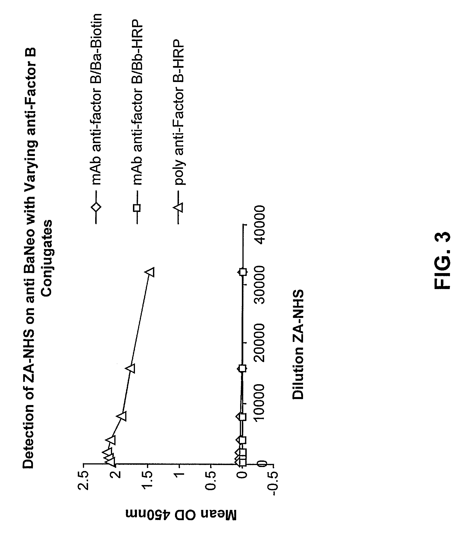
[0131]Nunc 96 well plates were coated with 10 μg / ml of the anti-Ba neo antibody overnight at 4° C. They were then incubated for one hour with a titration of zymosan activated normal human serum (ZA-NHS). The plate was subsequently washed with wash buffer and then incubated with biotinylated mAb anti Factor B / Ba, and mAb anti-Factor B / Bb-HRP as a control, and goat polyclonal Ab anti-Factor B-HRP. Excess conjugate was then removed, the biotinlyated mAb anti-Factor B / Ba was probed further with a streptavidin-horseradish peroxidase (SA-HRP) conjugate then washed and developed with 3,3′,5,5′-Tetramethylbenzidine (TMB) substrate and stopped with 1N HCl. See FIG. 3, showing that the antibody pair of mAb anti-human Ba neo and the polyclonal Factor B-HRP conjugated antibody are a highly sensitive matched pair and can detect Ba in a dilution series of ZA-NHS. The anti-Factor B-HRP is conjugated by Jackson Immunodiagnostics and is al...


PUM

PropertyMeasurementUnit
timeaaaaaaaaaa
timeaaaaaaaaaa
blood pressureaaaaaaaaaa

Abstract

Methods, compositions and kits for detecting the complement Factor B cleavage product Ba in a biological sample are described. These methods, compositions and kits are useful in convenient, reliable and early diagnosis of or ruling out pre-eclampsia in a pregnant human subject.

Description

CROSS-REFERENCE TO RELATED APPLICATIONS[0001]This application claims priority under 35 U.S.C. §119(e)(1) to U.S. Provisional Patent Application No. 61 / 500,574, filed 23 Jun. 2011, the contents of which are incorporated herein by reference in their entirety.REFERENCE TO SEQUENCE LISTING[0002]A “Sequence Listing” is submitted with this application in the form of a text file, created Sep. 7, 2012, and named “556408101 ST25.txt” (12,288 bytes), the contents of which are incorporated herein by reference in their entirety.TECHNICAL FIELD[0003]The present disclosure generally relates to the field of diagnostics, and, in particular, to a new technique for diagnosis in prenatal medicine, as well as methods and immunoassay kits for diagnosing, predicting or ruling out pre-eclampsia in a pregnant human subject.BACKGROUND[0004]The complement system is part of an innate system that provides an organism a natural defense against microbial agents and infection without the need for a specific antib...

Claims


Application Information

Patent Timeline
Patent Type & Authority Patents(United States)
IPC IPC(8): G01N33/53
CPCC07K16/18G01N2800/368G01N33/689G01N33/564C07K2317/33
Inventor DE FOREST, NIKOL LEETAMERIUS, JOHNNASSER, NOAHSEXTON, PATRICK
Owner QUIDEL
Learn More