Antibodies binding human collagen II
a technology of human collagen and antibodies, applied in the field of antibodies against human collagen ii, can solve the problems of abrasion of the articulating surface and adding a layer of complexity
- Summary
- Abstract
- Description
- Claims
- Application Information
AI Technical Summary
Benefits of technology
Problems solved by technology
Method used
Image
Examples
example 1
Identification of Collagen II Binding mAbs
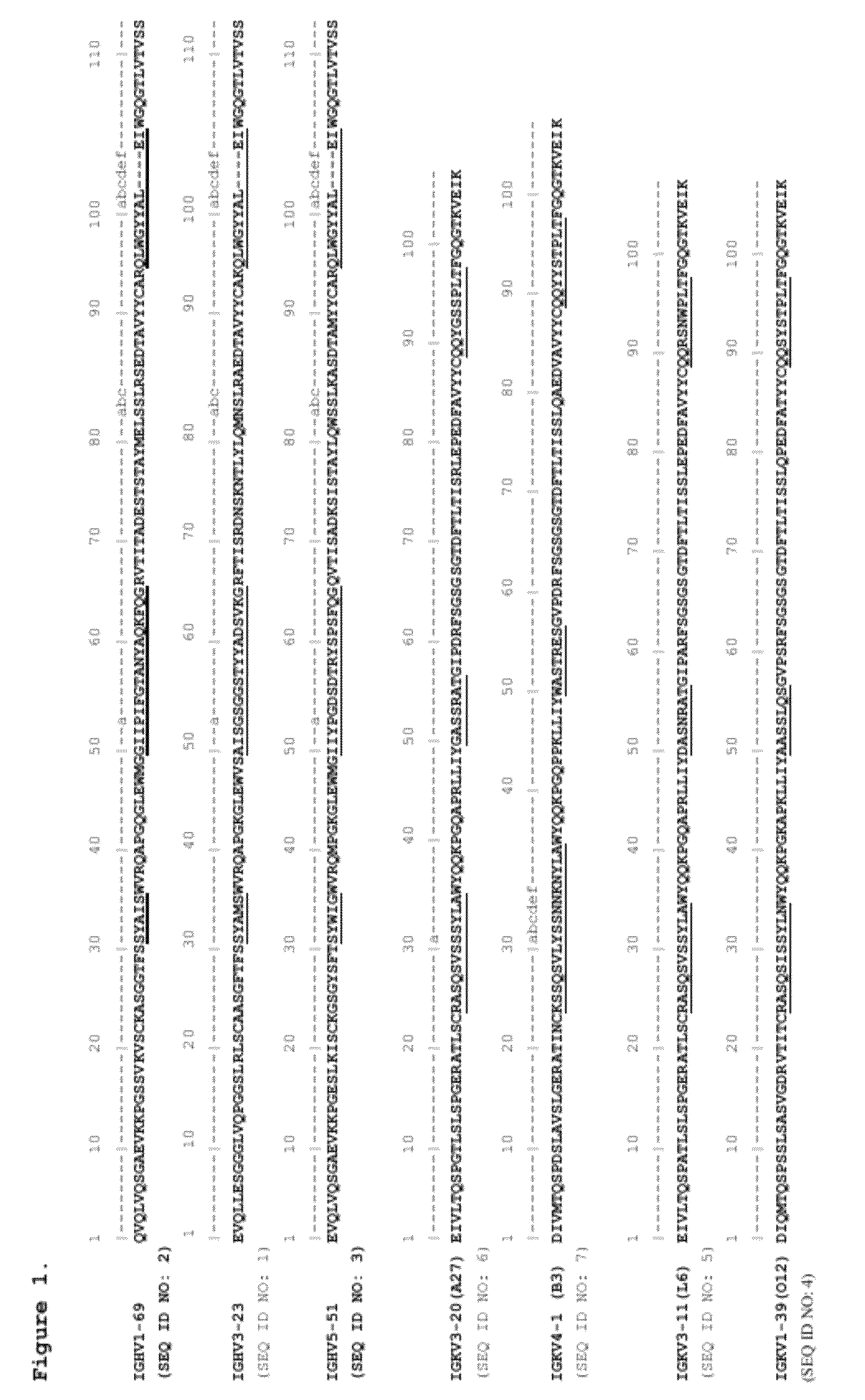
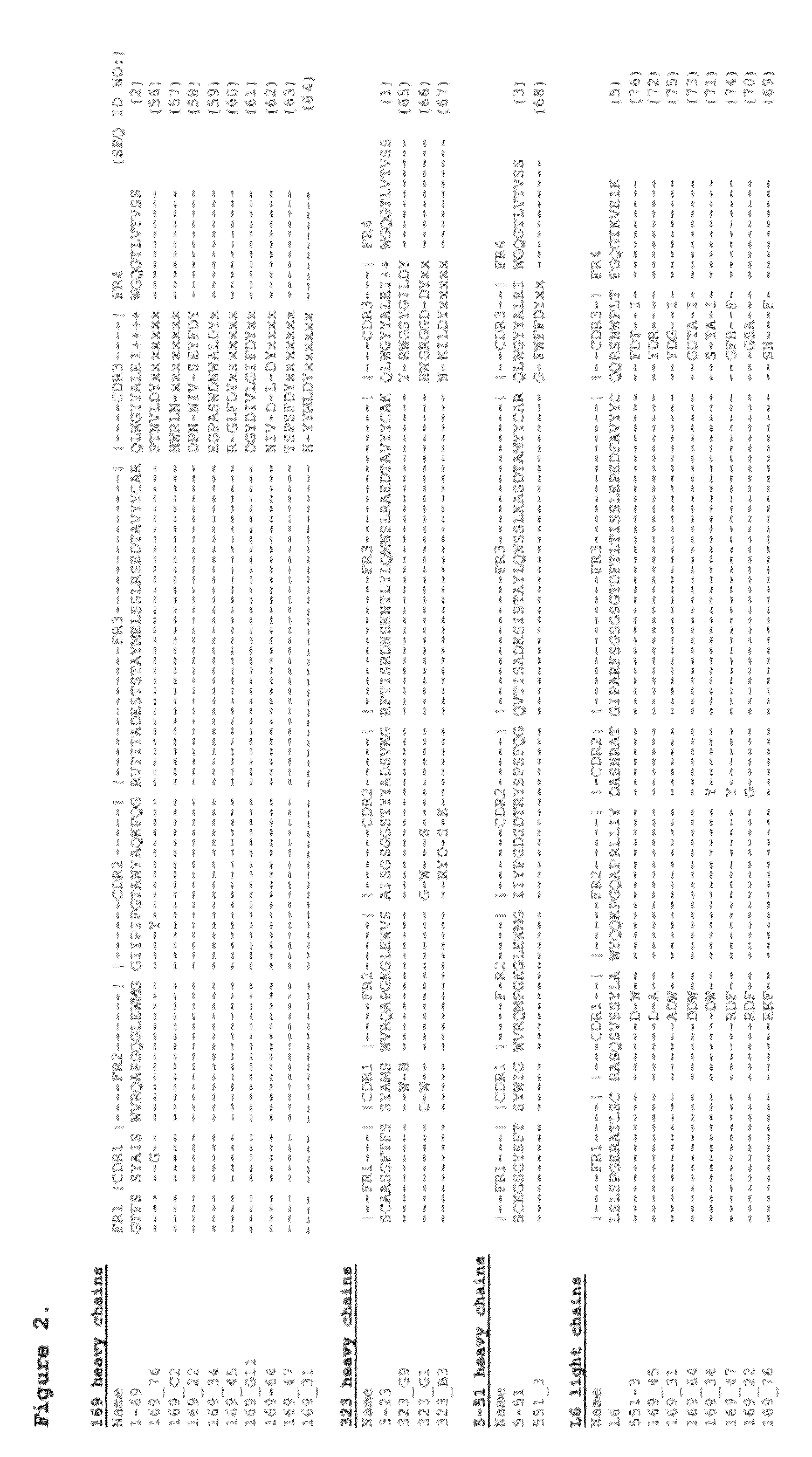
[0085]Human collagen II-binding Fabs were selected from de novo pIX phage display libraries (Shi et al., J. Mol. Biol. 397:385-396, 2010; WO2009085462A1; U.S. Pat. Appl. No. US20100021477). The libraries were generated by diversifying human germline VH genes IGHV1-69*01, IGHV3-23*01, and IGHV5-51*01, and human germline VLkappa genes 012 (IGKV1-39*01), L6 (IGKV3-11*01), A27 (IGKV3-20*01), and B3 (IGKV4-1*01). To assemble complete VH and VL domains, the IGHV genes were recombined with the human IGHJ-4 minigene via the H3 loop, and the IGKV genes were recombined with the IGKJ-1 minigene. The positions in the heavy and light chain variable regions around H1, H2, L1, L2 and L3 loops corresponding to positions identified to be frequently in contact with protein and peptide antigens were chosen for diversification. Sequence diversity at selected positions was limited to residues occurring at each position in the IGHV or IGLV germline gene families ...
example 2
Cross-Reactivity of Col II mAbs with Other Collagens
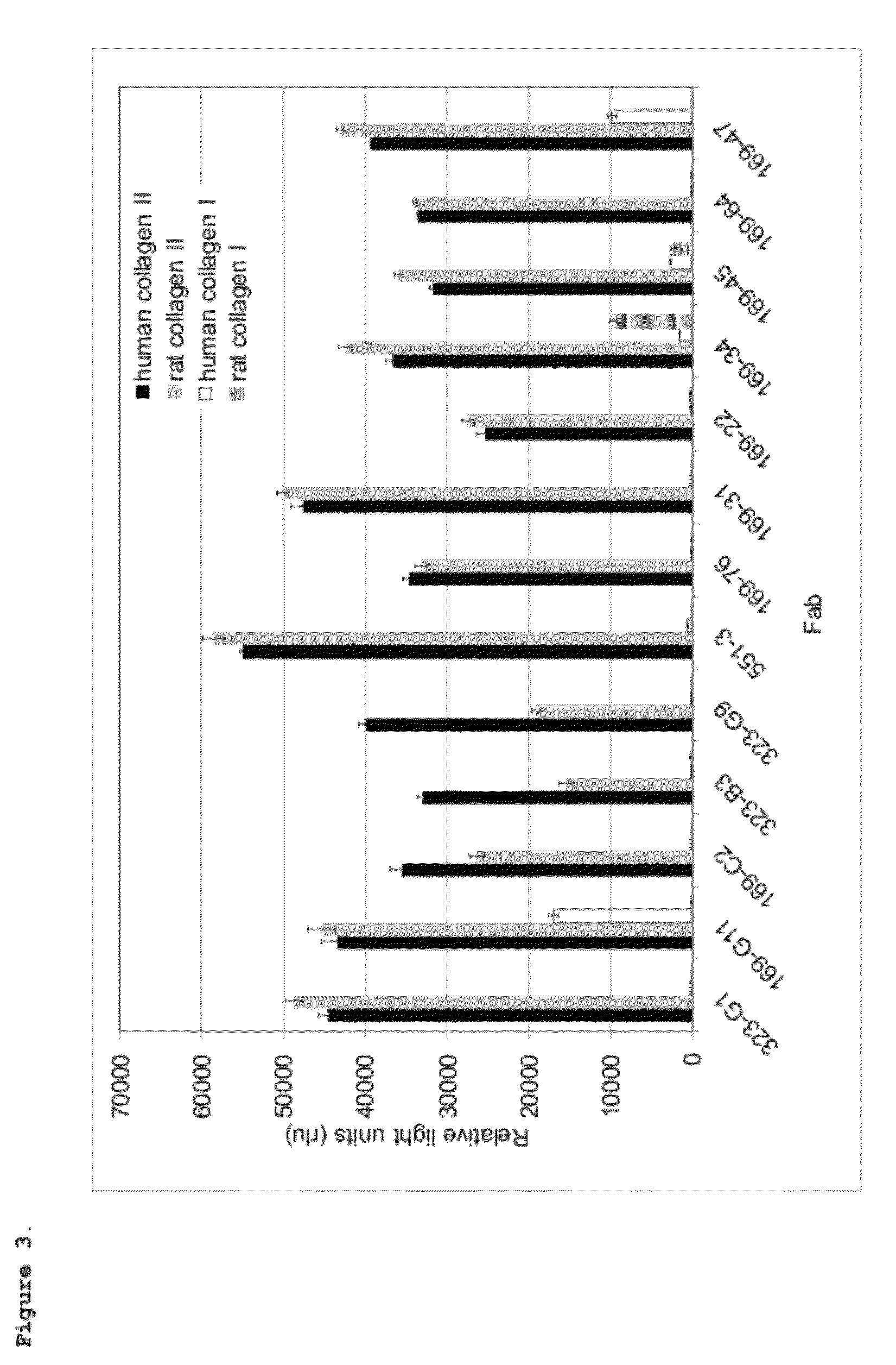
[0089]The human collagen II binding Fabs obtained from the initial panning were screened for cross-reactivity with human collagens I, IV and V, and rat collagens I and II.
Preparation of Fab Lysates.
[0090]Colonies were picked from the pIX-excised transformations and grown in 2xYT Carb. The next day, 50 μL of the saturated cultures were used to inoculate an expression plate containing 400 μL per well of 2xYT Carb and the plate was grown at 37° C. for 6 hours. Fab expression was induced with the addition of 1 mM IPTG and the plate was placed at 30° C. overnight. The next day, the induced Fab cultures were spun at 2000 rpm for 10 minutes, and the cleared lysate was used in subsequent assays.
ELISA
[0091]Maxisorp ELISA plates (Nunc) were coated with 10 μg / ml human collagen I (Chondrex), human collagen II (Chondrex), rat collagen I (Chondrex), rat collagen II (Chondrex) or with 5 μg / ml human collagen IV (Chemicon) or human collagen V (Chem...
example 3
Col II mAbs Bind to Human Cartilage
Small-Scale Fab Purification
[0092]The Fab expression in Escherichia coli in 2xYT Carb (except that TurboBroth (Athena ES) was used for expression of Fab 551-3) was induced with 1 mM IPTG at 30° C. overnight. Induced bacteria were pelleted 30 min 4500 rpm, the cell pellets resuspended in lysis buffer (20 mM Tris, pH 8.5, 350 mM NaCl, 7.5 mM imidazole) with protease inhibitors, and ruptured with two passes through a microfluidizer. The cell lysate was clarified with two spins at 10,000 rpm for 10 minutes. Talon resin (Clontech) was equilibrated with lysis buffer and two mLs were added to the clarified lysate. Bound Fabs were eluted with two incubations of 5 minutes each using elution buffer (150 mM EDTA, 20 mM Tris, pH 8.5) and dialyzed in 20 mM Tris, pH 8.5. The dialyzed Fabs were further purified using a Q-sepharose Fast Flow resin (QFF resin; GE Healthcare), and used for experiments.
[0093]Human cartilage was obtained from os...
PUM
| Property | Measurement | Unit |
|---|---|---|
| pH | aaaaa | aaaaa |
| temperature | aaaaa | aaaaa |
| time | aaaaa | aaaaa |
Abstract
Description
Claims
Application Information
 - R&D
- Intellectual Property
- Life Sciences
- Materials
- Tech Scout
- Unparalleled Data Quality
- Higher Quality Content
- 60% Fewer Hallucinations
Browse by: Latest US Patents, China's latest patents, Technical Efficacy Thesaurus, Application Domain, Technology Topic, Popular Technical Reports.
© 2025 PatSnap. All rights reserved.Legal|Privacy policy|Modern Slavery Act Transparency Statement|Sitemap|About US| Contact US: help@patsnap.com


