
Cardiac devices and methods for minimally invasive repair of ischemic mitral regurgitation

a technology of mitral valve and repair method, which is applied in the field of devices and methods for minimally invasive repair of mitral valve regurgitation, can solve the problems of ineffective annular reduction, increased surgical difficulty, and increased surgical difficulty, and achieves the effect of facilitating proper positioning of cutting instruments and/or monitoring the effectiveness of procedures

Active Publication Date: 2012-10-23
THE GENERAL HOSPITAL CORP
View PDF71 Cites 114 Cited by
  • Summary
  • Abstract
  • Description
  • Claims
  • Application Information

AI Technical Summary

Benefits of technology

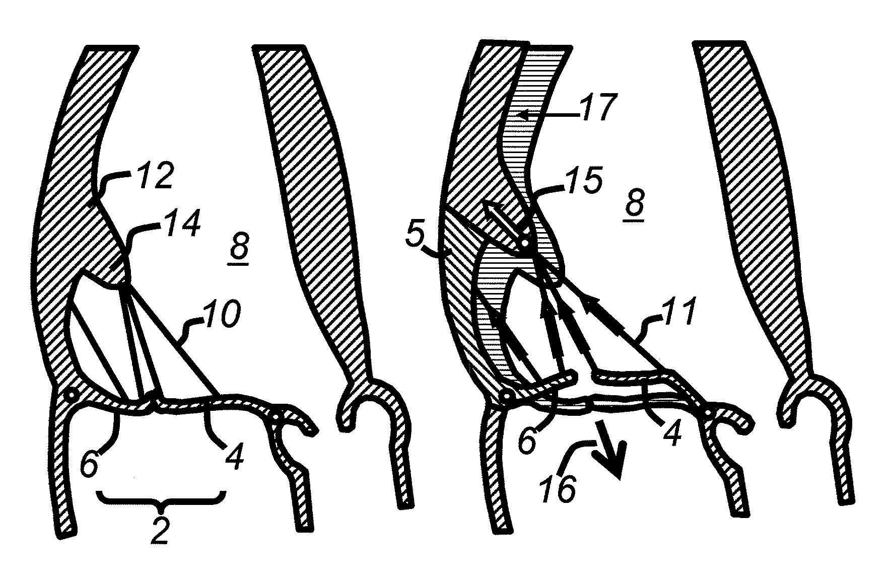
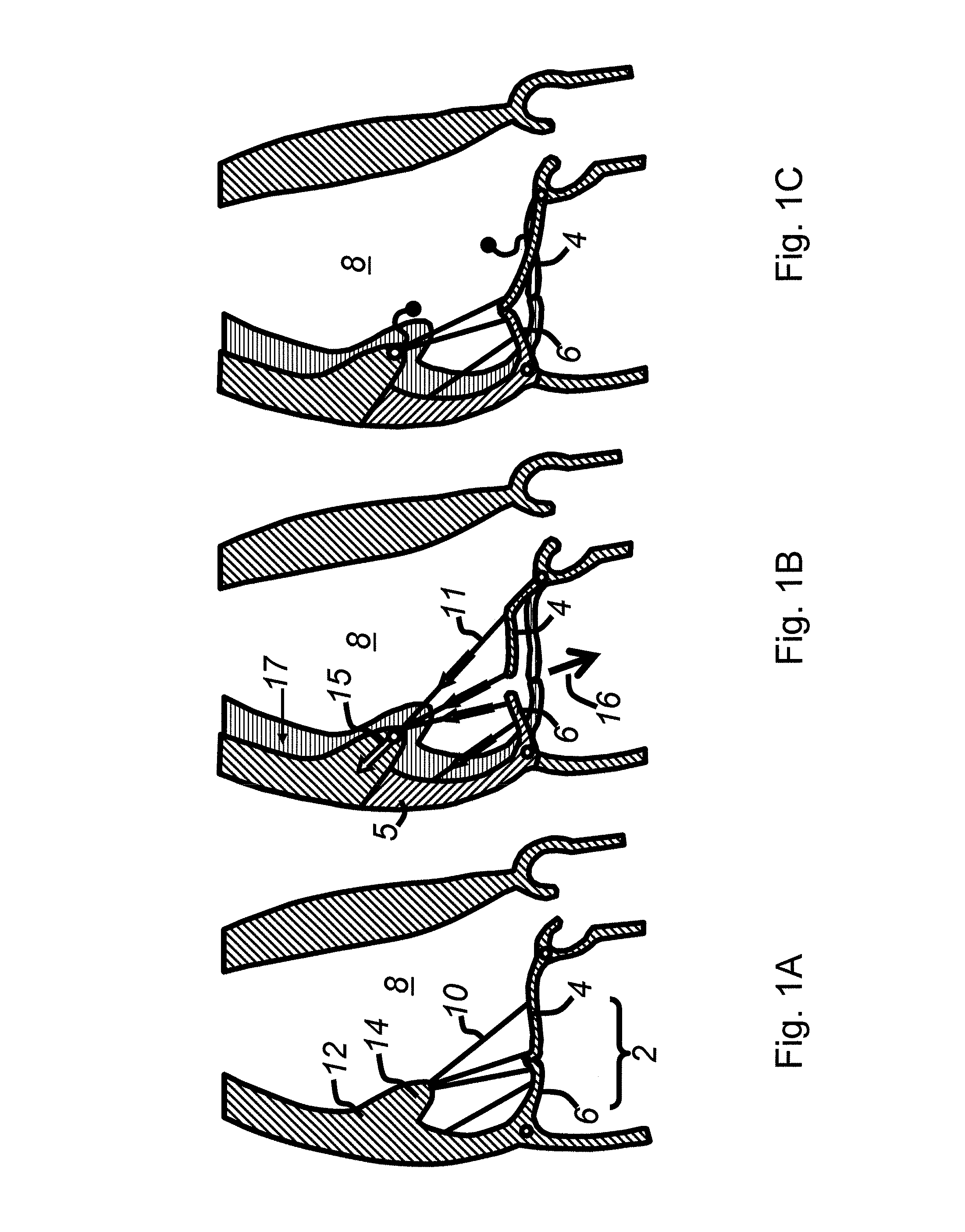
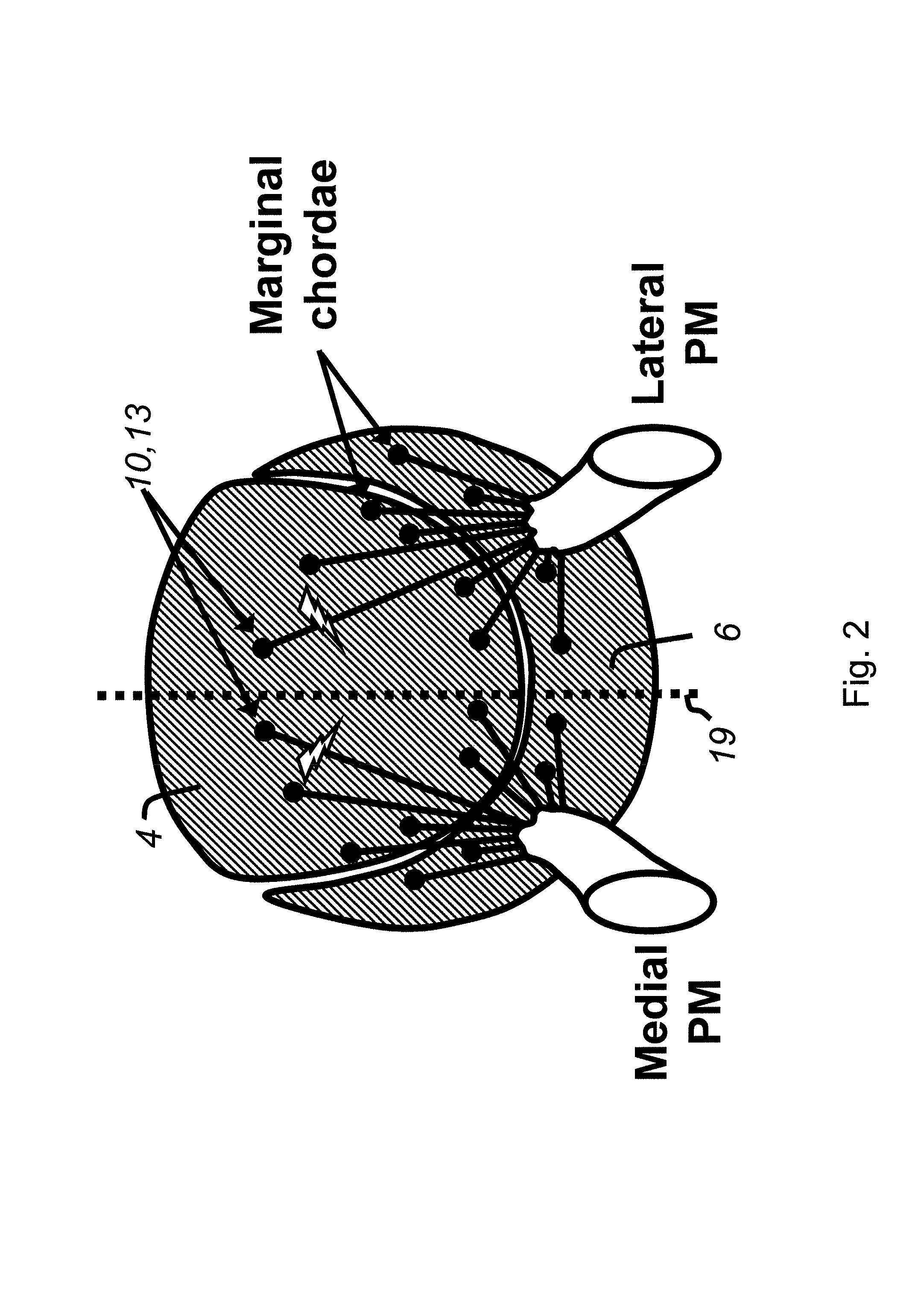
The present invention provides devices and methods for treating atrioventricular valve regurgitation by modifying the connection between the valve leaflets and the heart wall. This is achieved by severing one or more of the basal chords that attach the leaflets to the heart wall, which can cause bending of the leaflets and restrict their closure. The invention also involves elongating the chords by attaching two nodes to different points along their length and severing the chord between them. The technical effects of the invention include improved heart function, reduced heart size, and reduced risk of complications. The devices and methods are minimally invasive and can be performed using percutaneous catheters or imaging techniques.

Problems solved by technology

Such regurgitation limits exercise capacity by reducing forward blood flow to the body and overloading the lungs, causing fatigue and shortness of breath as cardinal manifestations of heart failure; if unrepaired, regurgitation also doubles the late mortality after heart attack and coronary artery bypass surgery.
Annular reduction, however, is often ineffective because it does not correct the fundamental heart wall deformation, so the mitral valve leaflets remain tethered.
Heart wall deformation or remodeling may also progress over time, so that even with initial success in reducing regurgitation, it often recurs after annular reduction therapy.
Moreover, in current practice, annular reduction procedures require putting patients on cardiopulmonary bypass (stopping the heart and opening it to insert the ring while using an artificial pump to bypass the heart and lungs), which itself conveys additional risk, deterring surgeons from performing this procedure.
Such techniques, however, require open-chest exposure of the heart and often extensive surgical manipulation.

Method used


Image

  • Cardiac devices and methods for minimally invasive repair of ischemic mitral regurgitation
  • Cardiac devices and methods for minimally invasive repair of ischemic mitral regurgitation
  • Cardiac devices and methods for minimally invasive repair of ischemic mitral regurgitation

Examples


Embodiment Construction

[0034]Preferred embodiments of the present invention will now be described with reference to the several figures of the drawing.

[0035]As shown in FIG. 3B, the present invention involves a percutaneous catheter 20 that may be advanced retrograde from the aorta into the outflow tract 22 of the left ventricle. The catheter is inserted using a standard guidewire approach (Seldinger technique), typically from the femoral artery in the groin. The catheter has a pre-formed or maneuverable bend that brings a cutting instrument 25 near a chord to be severed, such as the basal chord 10, which is the first chord encountered by catheter 20 just below the outflow tract, as seen in the ultrasound image pictured in FIG. 3A. (The tip of the catheter may be maneuvered, for example, using steering wires passed down its length.) In other embodiments, more than one chord will be cut. It may be preferable in some cases to sever pairs (symmetric or not) of chords, both of which are attached to the anteri...


PUM


Abstract

Novel apparatus and minimally invasive methods to treat atrioventricular valve regurgitation that is a result of tethering of chordae attaching atrioventricular valve leaflets to muscles of the heart, such as papillary muscles and muscles in the heart wall, thereby restricting the closure of the leaflets. Catheter embodiments for delivering and positioning chordal severing and elongating instruments are described.

Description

CROSS-REFERENCE TO RELATED APPLICATIONS[0001]This application claims priority of International Application No. PCT / US2003 / 024367 filed on Aug. 1, 2003, and U.S. Provisional Application No. 60 / 400,151 filed on Aug. 1, 2002, the entirety of which are incorporated herein by reference.FIELD OF THE INVENTION[0002]The present invention relates generally to devices (i.e., articles of manufacture, apparatus, systems, instruments) and methods for treating heart disease and, in particular, to devices and methods for minimally invasive repair of mitral valve regurgitation occurring in the context of cardiac pump dysfunction.BACKGROUND OF THE INVENTION[0003]With reference to FIG. 1A, the heart's mitral valve 2, comprised of an anterior leaflet 4 and a posterior leaflet 6, is the inlet valve to the main heart pumping chamber (left ventricle 8), and is forced to close when the ventricle contracts, preventing backward flow of blood. To ensure that this valve does not prolapse backward when the hea...

Claims


Application Information

Patent Timeline
Patent Type & Authority Patents(United States)
IPC IPC(8): A61B17/32A61B18/20A61BA61B17/00A61B17/22A61B17/28A61B17/3211A61B18/12A61B19/00A61F2/24A61M25/00
CPCA61B17/00234A61B17/221A61B18/24A61B18/1492A61B2017/00243A61B2017/003A61B2017/00349A61B2017/00867A61B2017/22035A61B2017/32004A61F2/2457
Inventor LEVINE, ROBERT A.MESSAS, EMMANUEL
Owner THE GENERAL HOSPITAL CORP
Learn More