
Activation of Egr-1 promoter by DNA damaging chemotherapeutics

a chemotherapeutic and promoter technology, applied in the field of egr1 promoter activation by dna damage chemotherapeutics, can solve the problems of ineffective chemotherapeutics used successfully to combat certain cancers, few chemotherapeutic agents are curative in human cancer treatment, and the subpopulation of cancerous cells to continue growing, so as to enhance the therapeutic response of neoplastics and avoid toxic effects

Active Publication Date: 2011-10-11
DANA FARBER CANCER INST INC +1
View PDF97 Cites 1 Cited by
  • Summary
  • Abstract
  • Description
  • Claims
  • Application Information

AI Technical Summary

Benefits of technology

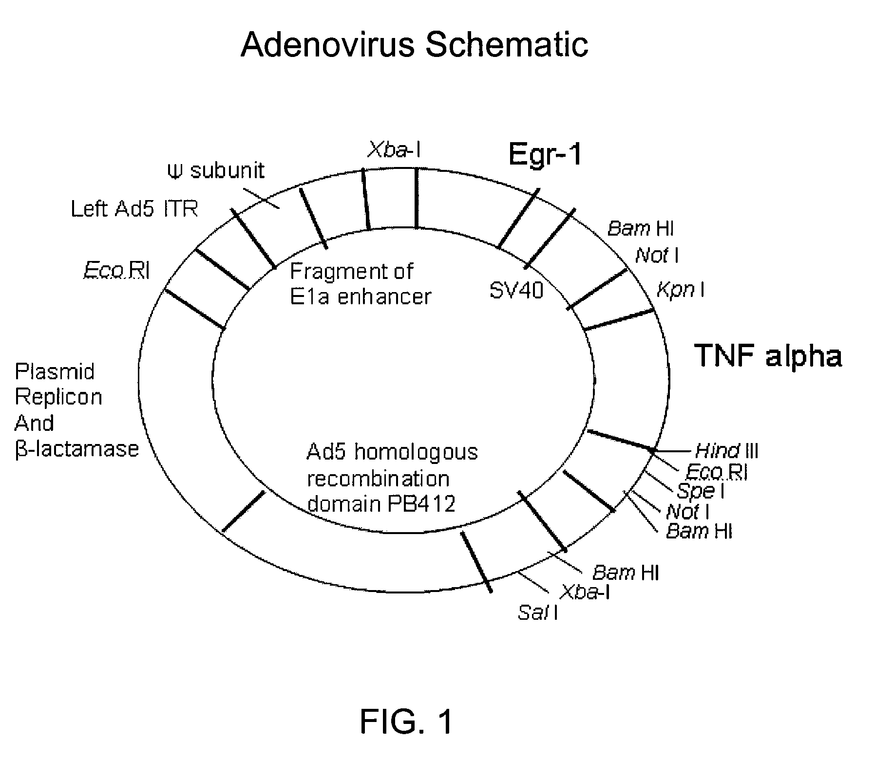
The present invention is about a method of delivering a therapeutic polypeptide to a subject using a chemotherapy agent. This method allows for targeted expression of the polypeptide, which can be toxic to cancer cells, and avoids the associated toxicity of systemic administration. The combined treatment of the therapeutic polypeptide with chemotherapy can enhance the therapeutic response of cancer cells. The invention also provides methods of inducing expression of a polypeptide in a cell and inhibiting the growth of a tumor in a subject. The expression of the therapeutic polypeptide is induced by a promoter operably linked to a polynucleotide encoding the polypeptide and the chemotherapy agent. The method can be used even in the presence of micrometastases. Overall, the invention provides a more effective and targeted approach to treat cancer.

Problems solved by technology

Chemotherapeutics that have been used successfully to combat certain cancers are frequently ineffective against other cancers, or are effective only at doses that are so high as to cause unacceptable toxicity.
Although cancer chemotherapy has advanced dramatically in recent years, very few chemotherapeutic agents are curative in human cancer treatment when delivered alone.
First, any single agent may only target a subset of the total population of malignant cells present, leaving a subpopulation of cancerous cells to continue growing.
Second, cells can develop resistance upon prolonged exposure to a drug.
However, the clinical effectiveness of cancer gene therapy has been limited by the lack of control of therapeutic gene expression within the tumor and selective targeting of the vector to the tumor.
In addition, certain combinations of agents may be synergistic; in other words, their combined effect is greater than predicted based on their individual activities.
However, combination therapies do not consistently provide the desired therapeutic effect and may contribute to multi-drug resistance.
In addition, antagonistic or biochemical interactions between the different therapies may lead to a reduced effectiveness and / or increased cytotoxicity for the combination than for either treatment alone.

Method used


Image

  • Activation of Egr-1 promoter by DNA damaging chemotherapeutics
  • Activation of Egr-1 promoter by DNA damaging chemotherapeutics
  • Activation of Egr-1 promoter by DNA damaging chemotherapeutics

Examples


example 1

Experimental Methods

[0071]The following materials and methods were used in the experiments described in Examples 2-5.

[0072]Cells and cell culture. Cell lines Seg-1, a human esophageal adenocarcinoma (Dr. David Beer, Univ. of Michigan, Ann Arbor, Mich.), and DHD / K12 / TRb (PROb), a rat colon adenocarcinoma established in syngeneic BD-IX rats by 1,2-dimethylhydrazine induction (Dr. Francois Martin, Univ. of Dijon, France), were maintained in Dulbecco's Modified Eagle Medium (DMEM) (GibcoBRL, Grand Island, N.Y.) supplemented with Fetal Bovine Serum (FBS, 10% v / v) (Intergen, Purchase, N.Y.), penicillin (100 IU / ml), and streptomycin (100 μg / ml) (GibcoBRL), at 37° C. and 7.5% CO2.

[0073]Animals. Athymic nude mice (Frederick Cancer Research Institute, Frederick, Md.) received food and water ad libitum. Experiments were in accordance with the guidelines of the University of Chicago.

[0074]Viral vectors. The viral vectors Ad.Egr.TNF.11D and Ad.Null.3511.11D (GenVec, Gaithersburg, Md.) were store...

example 2

In vitro Induction of TNF-α in Human and Rat Tumor Cells Following Infection with Ad.Egr.TNF.11D and Exposure to Cisplatin

[0081]TNF-α production was tested in human esophageal Seg-1 cells and rat colorectal PROb cells infected with Ad.Egr.TNF.11D or Ad.Null.3511.11D following exposure to IR or cisplatin, using an ELISA specific for human TNF-α.

[0082]No TNF-α protein was detectable in Seg-1 pellets or supernatants from cultures infected with Ad.Null.3511.11D and treated with IR or cisplatin, moderate levels were detected in cells infected with Ad.Egr.TNF.11D alone (269.3±1.9, 167.8±8.4, 260.6±14.9; P<0.001), and significant levels were detected in cells infected with Ad.Egr.TNF.11D and exposed to 5 Gy IR (768.8±32.6, 593.0±27.6, 746.0±18.5), and to 5 μM cisplatin (885.3±28.7, 892.6±21.3, 901.7±21.7; P<0.001), at 24, 48 and 72 hours, respectively. Combined treatment of Ad.Egr.TNF.11D and IR resulted in a 2.9, 3.5 and 2.9-fold increase, and Ad.Egr.TNF.11D and cisplatin resulted in a 3....

example 3

CArG Elements of the Egr-1 Promoter Mediate Induction of TNF-α by Cisplatin

[0085]Seg-1 and PROb cells were co-transfected with the firefly luciferase reporter plasmid constructs pGL3 basic (negative control), pGL3 660 (minimal Egr-1 promoter, no CArG elements), or pGL3 425 (all CArG elements, no AP-1 sites), and the Renilla luciferase reporter plasmid construct pRL-TK.

[0086]Luciferase activity (LA) was minimal in Seg-1 cells transfected with the pGL3 basic (0.01-0.02) and the pGL3 660 (0.10-0.18) plasmid constructs, while cells transfected with the pGL3 425 plasmid construct exhibited a 2.4-fold increase in relative LA (15.07) following exposure to 20 Gy IR compared to untreated control (6.37) and a 2.0-fold increase in LA (2.89) following exposure to 50 μM cisplatin compared with untreated control. See FIG. 7A. Similarly, LA was minimal in PROb cells transfected with the pGL3 basic (0.21-0.30) and the pGL3 660 (0.76-1.84) plasmid constructs, while cells transfected with the pGL3 42...


PUM

PropertyMeasurementUnit
timeaaaaaaaaaa
pHaaaaaaaaaa
pHaaaaaaaaaa

Abstract

The present invention relates to methods of inducing expression of a polynucleotide encoding a therapeutic polypeptide, e.g., TNF-α, in a cell comprising contacting the cell with a construct comprising an Egr-1 promoter operably linked to a polynucleotide encoding the polypeptide, and at least one chemotherapeutic agent, wherein the chemotherapeutic agent induces expression of the polypeptide. The invention also relates to methods of inhibiting a neoplastic cell, comprising contacting the cell with a construct comprising an Egr-1 promoter operably linked to a polynucleotide encoding TNF-α and a chemotherapeutic agent. The present invention further relates to methods of inhibiting or reducing the growth of a tumor in a subject, comprising co-administering to the subject a construct comprising an Egr-1 promoter operably linked to a polynucleotide encoding TNF-α and a chemotherapeutic agent, wherein the co-administration inhibits or reduces the ability of the tumor to grow.

Description

CROSS REFERENCE TO RELATED APPLICATIONS[0001]This application is a continuation-in-part of U.S. application Ser. No. 10 / 117,442, filed Apr. 5, 2002, now abandoned, which claims priority to U.S. Provisional Application No. 60 / 282,040, filed Apr. 6, 2001, and is also a continuation-in-part of U.S. application Ser. No. 10 / 795,090, filed Mar. 5, 2004, now abandoned, which claims priority to U.S. Provisional Application No. 60 / 452,489, filed Mar. 6, 2003. The entire texts of the above-referenced disclosures are hereby incorporated by reference.INTRODUCTION[0002]Therapy for cancer has largely involved the use of chemotherapy, in which highly toxic chemicals are given to the patient, and / or radiotherapy, in which toxic doses of radiation are administered. Chemotherapeutics that have been used successfully to combat certain cancers are frequently ineffective against other cancers, or are effective only at doses that are so high as to cause unacceptable toxicity. Although cancer chemotherapy...

Claims


Application Information

Patent Timeline
Patent Type & Authority Patents(United States)
IPC IPC(8): A61K48/00C12N5/07A01N65/00A01N63/00C12N5/10C07H21/04C07H21/02A61K33/243
CPCA61K31/66A61K31/704A61K31/7072A61K33/24A61K38/191A61K45/06A61K2300/00A61K33/243
Inventor WEICHSELBAUM, RALPH R.KUFE, DONALD W.POSNER, MITCHELLMAUCERI, HELENAPARK, JAMES O.
Owner DANA FARBER CANCER INST INC
Learn More