Method for screening for vitamin D receptor ligands
a technology of vitamin d receptor and ligand, which is applied in the field of screening for compounds participating in osteoblast differentiation, can solve the problems of unclear mechanism of bone mass increase mediated by vitamin dsub>3/sub>derivatives, poor effect of increasing the bone density of lumbar vertebrae or collum femoris, etc., and achieves increased osteoblast differentiation, increased vdr ligand-dependent transcription activity, and enhanced activity
- Summary
- Abstract
- Description
- Claims
- Application Information
AI Technical Summary
Benefits of technology
Problems solved by technology
Method used
Image
Examples
example 1
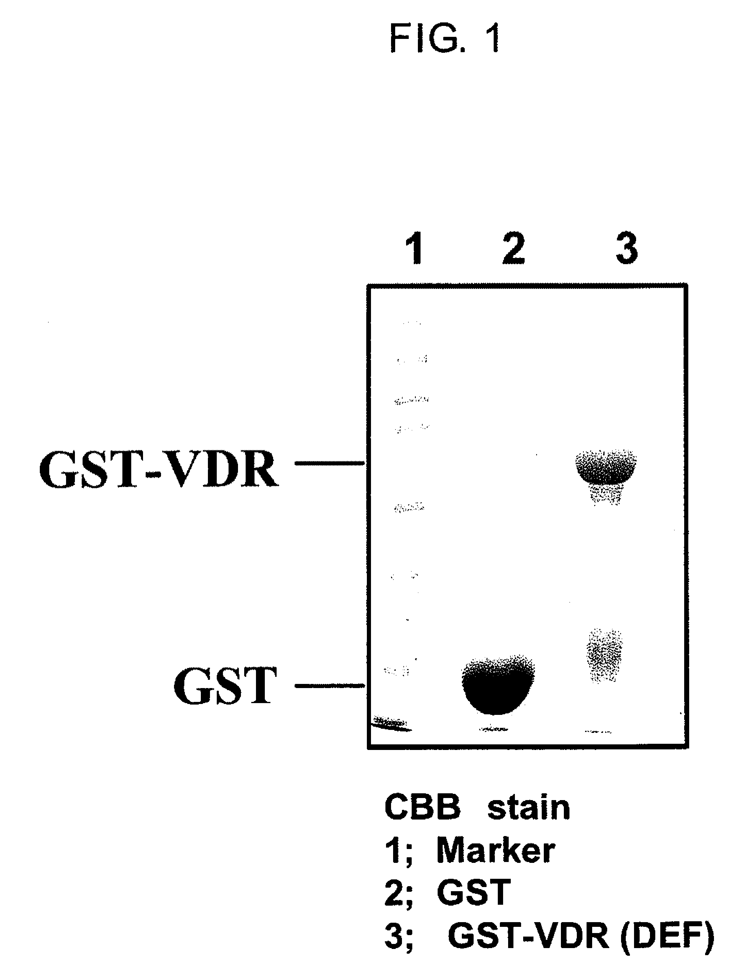
Purification of Protein Complex of Fusion Proteins Between VDR and Glutathione-S-Transferase (GST)
[0117]Firstly, GST-VDR which is a bait protein was expressed in E coli. With respect to rat VDR sequences reported by Kitagawa et al., Cell, Vol. 113, 905-917, 2003, E coli. (DH5alpha strain) transformed using either a pGEX4T-1 vector containing the ligand binding region (Amersham Biosciences) or a pGEX4T-1 vector containing DNA sequences encoding the human VDR ligand binding region (DEF region) (The structure of the VDR domain is explained in J Wesley Pike et al. The Vitamin D Receptor. (2005) Vitamin D, 2nd Edition, Feldman, Pike, Glorieux (eds.) p. 167-191) was cultured in 2 L of LB medium at 37° C., while shaking at 120 revolutions. When the absorbance at 600 nm reached 0.4-0.5, 0.1 mM isopropyl-1-thio-β-D-galactopyranoside (IPTG) was added to the culture and culturing was continued for 3 hours at 27° C. while shaking at 120 revolutions in order to induce GST-VDR protein expression....
example 2
Identification and Purification of Protein Bound to GST-VDR (DEF)
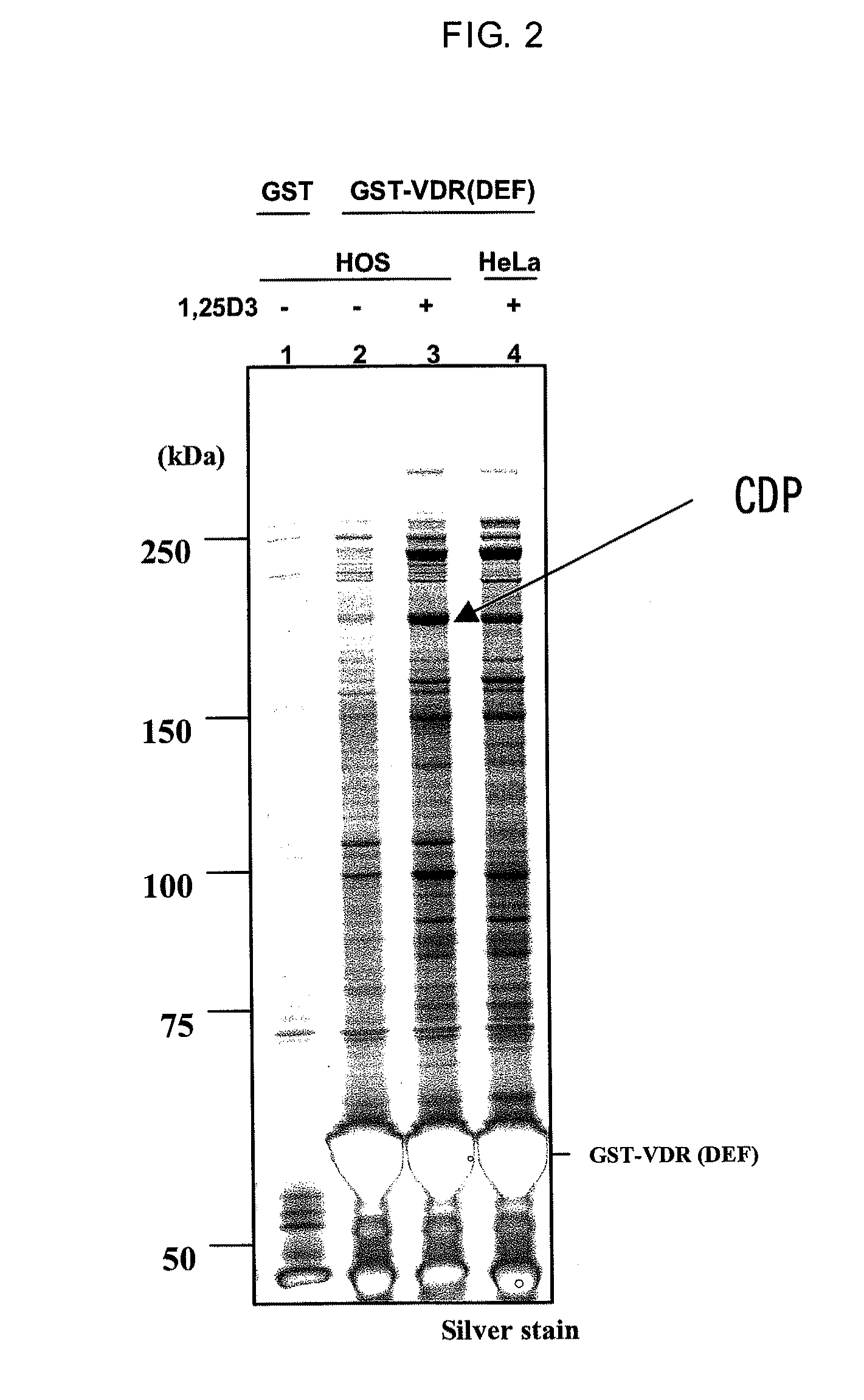
[0118]In accordance with a method described in Dignam J D. et al., Nucleic Acids Research, 1983, Vol. 11, No. 5 1475-1489, a nuclear protein extract was prepared from HOS cells which is a human osteoblast cell strain (obtained from ATCC, Number CRL-1543). The resulting nuclear protein extract was dissolved in buffer D (20 mM HEPES, 10% glycerol, 0.15 M KCl, 0.2 mM EDTA, 0.5 mM PMSF, 0.01% 2-mercaptoethanol (pH=7.9). The nuclear protein extract was mixed for one hour or overnight with GST-VDR resin either in the presence or absence of 1,25(OH)2D3 which is a VDR ligand. The GST-VDR resin was washed and placed in a state in which only a specifically binding protein was bound to the GST-VDR resin. The protein bound to the resin was eluted using a reduced glutathione solution (pH=8.0) having a concentration of 15 mM on the GST-VDR resin. The eluted protein was subjected to SDS-PAGE under reducing conditions in the same mann...
example 3
Detection of CDP by Western Blotting
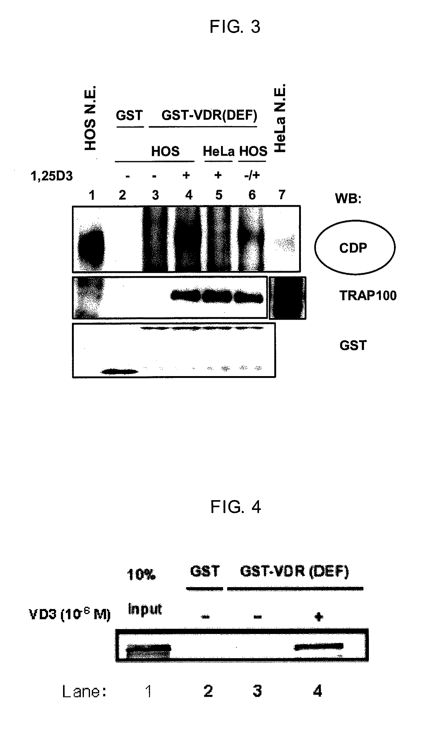
[0119]A part of the eluted liquid from Example 2 was subjected to SDS-PAGE under reducing conditions, blotted to a PVDF membrane (Millipore, Product No. IPVH304F0) and the detection of CDP was performed using standard techniques. The primary antibodies were anti-CDP (M-222) rabbit polyclonal IgG (Santa Cruz Biotechnology, Product No. sc-13024). The secondary antibody was anti-rabbit IgG labeled with HRP (Amersham Biosciences, Product No. NA934V). ECL Plus (GE Healthcare Bioscience, Product No. RPN2132) was used in the detection operation. The results are shown in FIG. 3. Although a CDP band was not detected nearly at all in Lane 3 in the absence of a ligand, a CDP band was strongly detected in Lane 4 in the presence of a ligand. For comparison, a nuclear protein extract from HeLa cells which are uterine cancer-derived cells rather than osteoblasts was obtained in the same manner and mixed with GST-VDR in the presence of a ligand. The elution of th...
PUM
| Property | Measurement | Unit |
|---|---|---|
| excitation wavelengths | aaaaa | aaaaa |
| excitation wavelengths | aaaaa | aaaaa |
| excitation wavelengths | aaaaa | aaaaa |
Abstract
Description
Claims
Application Information
 - R&D
- Intellectual Property
- Life Sciences
- Materials
- Tech Scout
- Unparalleled Data Quality
- Higher Quality Content
- 60% Fewer Hallucinations
Browse by: Latest US Patents, China's latest patents, Technical Efficacy Thesaurus, Application Domain, Technology Topic, Popular Technical Reports.
© 2025 PatSnap. All rights reserved.Legal|Privacy policy|Modern Slavery Act Transparency Statement|Sitemap|About US| Contact US: help@patsnap.com


