Lipid-drug conjugates for local therapy of eye diseases
- Summary
- Abstract
- Description
- Claims
- Application Information
AI Technical Summary
Benefits of technology
Problems solved by technology
Method used
Image
Examples
example 1
Synthesis of Compounds
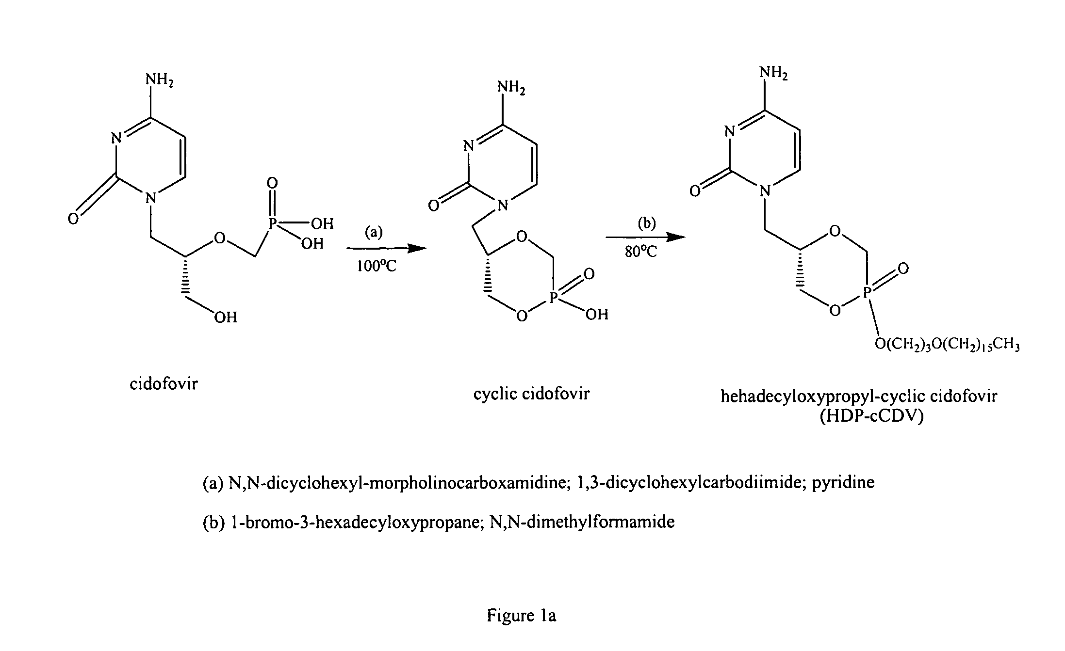
[0080]Hexadecylpropanediol-3-phospho-ganciclovir (HDP-P-GCV) was synthesized as previously reported.2 To prepare a small particle formulation and eliminate the population of large particles, HDP-P-GCV was suspended in distilled water and the slurry was subjected to five passes through a microfluidizer (Microfluidics, Newton, Mass.). The slurry was then flash frozen in a 1 liter round bottom flask and lyophilized overnight to remove the water.
[0081]Unmodified HDP-P-GCV and microfluidized HDP-P-GCV were subjected to laser light scattering particle size analysis at Cirrus Pharmaceuticals, Inc., Durham, N.C. Measurements were performed using a HELOS laser diffraction instrument (Sympatec, Lawrenceville, N.J.), equipped with a R3 lens (0.5 to 175 microns).
[0082]For each measurement, approximately 100 mg of dry sample was dispersed at a feed rate of 75% using the VIBRI and RODOS attachments set at a main pressure of 4.0 bar and aventury pressure of 100 mbar. The trig...
example 2
Animal Studies
[0083]All procedures were adherent to the ARVO Statement for the Use of Animals in Ophthalmic and Vision Research Intravitreal pharmacokinetics of small particle formulation of HDP-P-GCV: Three rabbits received 8.85 μmoles of the drug in their right eyes and 2.8 μmoles in the left eye. 2.8 μmole was previously determined to be non toxic.2 Vitreous sampling was performed on both eyes of each rabbit at post injection week 1, week 2, week 3, week 5, week 8, week 12, and week 15. 50 to 100 μl of vitreous fluid was aspirated and placed in a pre-weighed vial for HPLC analysis of HDP-P-GCV levels as described before. 2. Intravitreal toxicity and pharmacokinetics of HDP-cCDV: For toxicity studies, five doses (10 μg or 0.018 μmole, 55 μg or 0.1 μmole, 100 μg or 0.18 μmole, 550 μg or 1.01 μmole, 1000 μg or 1.84 μmole in 50 μl of 5% dextrose) were tested in 8 rabbits, 16 eyes for 8 weeks. One eye of each animal was injected with drug and the fellow eye was injected with 5% dextro...
example 3
Effect of Microfluidization of HDP-P-GCV on Particle Size
[0086]An aqueous slurry of HDP-P-GCV was subjected to five consecutive cycles of microfluidization. After lyophilization and recovery of the powder, both the unmodified and the microfluidized HDP-P-GCV formulations were subjected to laser light scattering particle size analysis (FIG. 3). Unmodified HDP-P-GCV (Panel A) showed a bimodal distribution with a volume median diameter (×50) centered around 8.0 microns. After microfluidization (Panel B), a more monodispersed population of smaller particles is noted, having a ×50 of 4.4 microns and a 99th percentile diameter (×99) of 20 microns and a 90th percentile diameter (×90) of 10 microns. No large particles remained after microfluidization treatment. Finally, an untreated formulation of HDP-cCDV powder (Panel C) was also analyzed. This compound showed a population of particles having a ×50 of 8.9 microns.
[0087]Intravitreal pharmacokinetics of the unmodified and microfluidized sma...
PUM
| Property | Measurement | Unit |
|---|---|---|
| Time | aaaaa | aaaaa |
| Nanoscale particle size | aaaaa | aaaaa |
| Nanoscale particle size | aaaaa | aaaaa |
Abstract
Description
Claims
Application Information
 - R&D
- Intellectual Property
- Life Sciences
- Materials
- Tech Scout
- Unparalleled Data Quality
- Higher Quality Content
- 60% Fewer Hallucinations
Browse by: Latest US Patents, China's latest patents, Technical Efficacy Thesaurus, Application Domain, Technology Topic, Popular Technical Reports.
© 2025 PatSnap. All rights reserved.Legal|Privacy policy|Modern Slavery Act Transparency Statement|Sitemap|About US| Contact US: help@patsnap.com


