
Method for quantification of methylated DNA

a methyl cytosine and dna technology, applied in the field of methyl cytosine quantification in dna, can solve the problems of pcr amplification, inapplicability of usual methods for identifying nucleic acids, and loss of epigenetic information carried by 5-methylcytosine during pcr amplification

Inactive Publication Date: 2011-07-05
EPIGENOMICS AG
View PDF5 Cites 6 Cited by
  • Summary
  • Abstract
  • Description
  • Claims
  • Application Information

AI Technical Summary

Benefits of technology

This method provides improved accuracy and reliability in quantifying methylated DNA, reducing errors and biases associated with traditional endpoint analysis, and enhances the sensitivity and specificity of cancer diagnostics by accurately measuring methylation levels in bodily fluids.

Problems solved by technology

However, a detection of 5-methylcytosine is difficult because 5-methylcytosine has the same base pairing behaviour as cytosine.
Therefore the usual methods for identifying nucleic acids are not applicable.
Moreover, the epigenetic information carried by 5-methylcytosine is completely lost during PCR amplification.
However, a reliable diagnosis by detecting specially methylated cytosines in body fluids is difficult, because the aberrant methylation pattern has to be found within a large amount of background DNA, which is methylated differently, but which has the same base sequence.
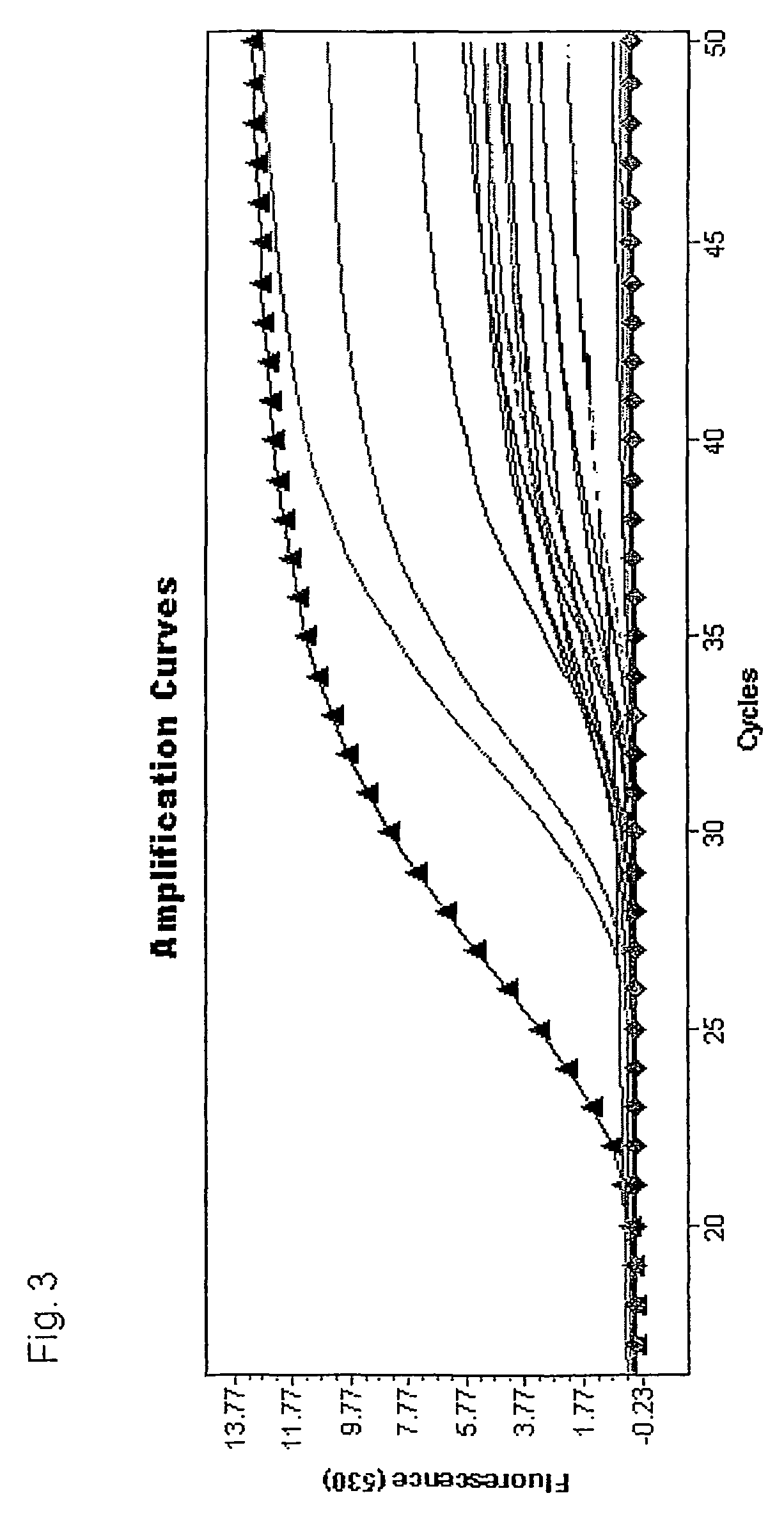
However, this “endpoint analysis” leads to several problems: due to product inhibition, enzyme instability and decrease of the reaction components the amplification does not proceed uniformly.
As a consequence, the quantification is error-prone (for review: Kains: The PCR plateau phase—towards an understanding of its limitations.
However, both methods do not produce exact results for the quantification of methylation via MethyLight assays.
These downscaled curves are the reason that both analysis methods generate incorrect results.
However, for samples with low proportions of DNA that is methylated at the probe (especially common in cancer diagnostics) this is not true.

Method used


Image

  • Method for quantification of methylated DNA
  • Method for quantification of methylated DNA
  • Method for quantification of methylated DNA

Examples


example 2

[0042]In this experiment 1.8 mL serum samples from 30 patients without colon cancer and 30 patients with colon cancer or colon polyps were measured using a MSP MethyLight assay with hydrolysis (Taqman™) probes. The aim is to predict the presence of colon cancer with high sensitivity and specificity. The area under the ROC curve (AUC) is used to quantify the prediction performance. A Receiver Operating Characteristic (ROC) curve is a plot of the true positive rate against the false positive rate for the different possible cutpoints of a diagnostic test. It shows the tradeoff between sensitivity and specificity depending on the selected cutpoint (any increase in sensitivity will be accompanied by a decrease in specificity). The area under an ROC curve (AUC) is a measure for the accuracy of a diagnostic test (the larger the area the better, optimum is 1, a random test would have a ROC curve lying on the diagonal with an area of 0.5; for reference: J. P. Egan. Signal Detection Theory an...


PUM

PropertyMeasurementUnit
real time PCRaaaaaaaaaa
shapeaaaaaaaaaa
real timeaaaaaaaaaa

Abstract

The present invention relates to a method for the quantification of methylated cytosines in DNA. In the first step of the invention unmethylated cytosines in the DNA to be analysed are chemically converted into uracil while 5-methylcytosines remain unchanged. In a second step the converted DNA is amplified methylation specifically in a real time PCR using a methylation specific probe. Finally the amount of uniformly methylated DNA is calculated by combining criteria derived from the shape of the real time curve and from the signal intensity. The method is preferably used for diagnosis and / or prognosis of adverse events for individuals, for distinguishing cell types and tissues, or for investigating cell differentiation.

Description

BACKGROUND OF THE INVENTION[0001]The present invention relates to a method for the quantification of methylated cytosines in DNA. 5-methylcytosine is the most frequent covalent base modification in the DNA of eukaryotic cells. It plays an important biological role, e.g. in the regulation of the transcription, in genetic imprinting, and in tumorigenesis (for review: Millar et al.: Five not four: History and significance of the fifth base. In: The Epigenome, S. Beck and A. Olek (eds.), Wiley-VCH Verlag Weinheim 2003, p. 3-20). Therefore, the identification of 5-methylcytosine as a component of genetic information is of considerable interest. However, a detection of 5-methylcytosine is difficult because 5-methylcytosine has the same base pairing behaviour as cytosine. Therefore the usual methods for identifying nucleic acids are not applicable. Moreover, the epigenetic information carried by 5-methylcytosine is completely lost during PCR amplification.[0002]The usual methods for methyl...

Claims


Application Information

Patent Timeline
Patent Type & Authority Patents(United States)
IPC IPC(8): C12Q1/68C12P19/34
CPCC12Q1/6858Y10T436/143333C12Q2561/101C12Q2535/125C12Q2523/125C12Q2525/186
Inventor MODEL, FABIAN
Owner EPIGENOMICS AG
Learn More