
Quadruple inversion recovery for quantitative contrast-enhanced black blood imaging

a black blood imaging and quadriple inversion technology, applied in the field of magnetic resonance imaging (mri), can solve problems such as providing efficient suppression

Inactive Publication Date: 2010-05-11
UNIV OF WASHINGTON
View PDF12 Cites 3 Cited by
  • Summary
  • Abstract
  • Description
  • Claims
  • Application Information

AI Technical Summary

Benefits of technology

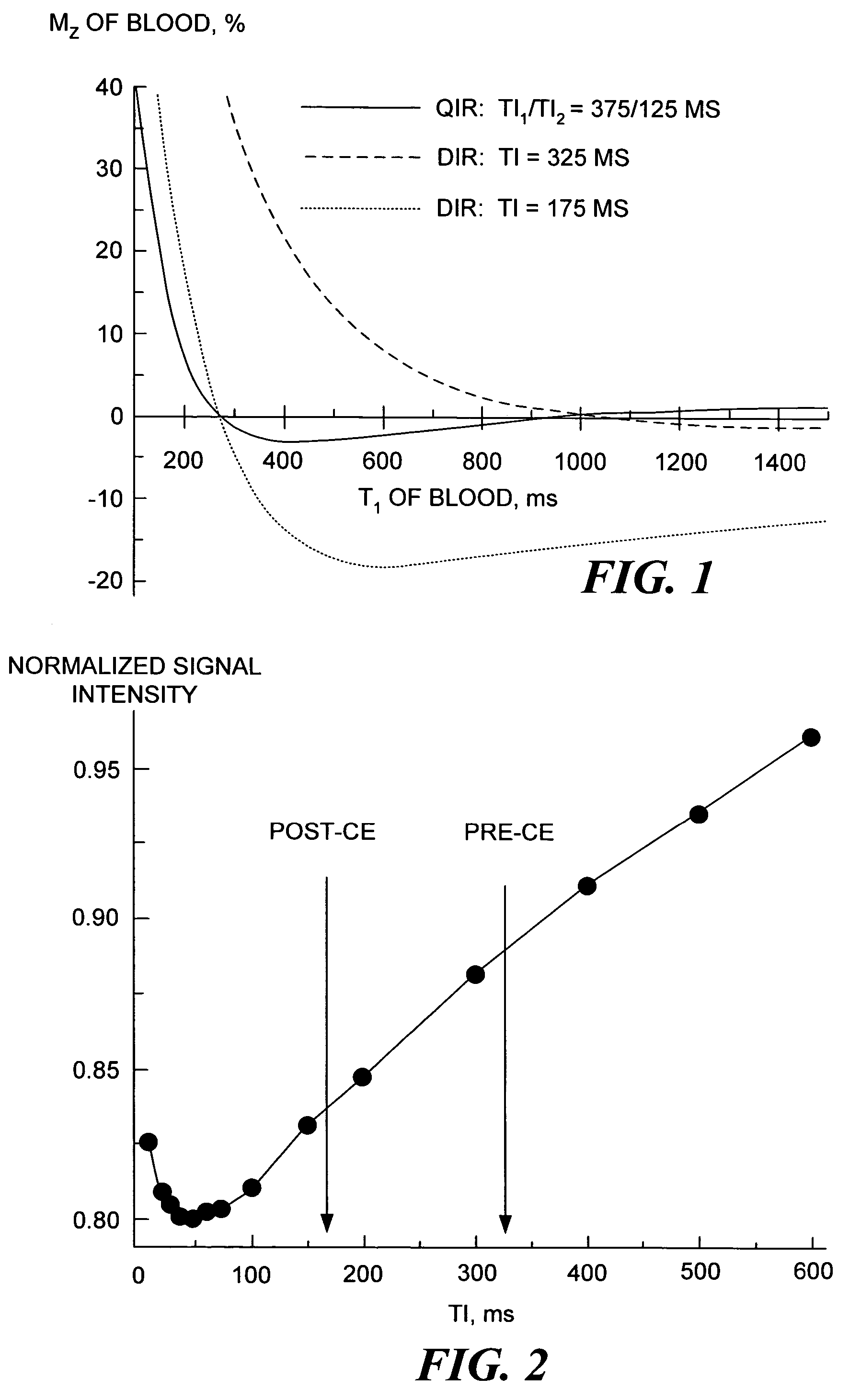
[0005]As explained above, the current approach used for black-blood imaging in MRI of the heart and blood vessels has typically applied the DIR method. However, DIR only provides efficient suppression of the blood signal within a narrow range of T1 values at any selected TI. The present invention addresses this issue by suppressing the signal and artifacts from flowing blood in MRI of the heart and blood vessels over a wide range of T1 values, so that the changes in T1 caused by the infusion of a CE agent are not problematic in achieving high-quality black blood images.
[0007]The longitudinal magnetization of the blood varies as a combination of exponential functions of several parameters. These parameters include the first predefined inversion delay period, the second predefined inversion delay period, a repetition time, TR, and a current T1 value of the blood, which depends on the concentration of the CE agent infused. The CE agent reduces the T1 value for blood in an unpredictable manner. However, the residual magnetization of the blood is negligibly small for a wide range of T1 after application of the sequence of RF pulses in accord with the present invention. This range covers all possible T1 variability. Thus, the method produces consistently black-blood images independently of the presence and concentration of the CE agent in the blood.
[0008]In another aspect of the invention, a method for black-blood contrast enhanced MRI is defined. The method starts with the step of infusing the CE agent into the blood. The CE agent used preferably includes gadolinium derivatives, although other suitable CE agents may instead be used. After waiting a sufficient time, blood carrying a portion of the CE agent flows through the site. At that time, the images are acquired using the method of the present invention, which includes two double-inversion procedures, each of which is followed by one of two predefined time periods. As was noted above, this method effectively suppresses the blood signal. Thus, tissues surrounding blood are clearly visible after CE without any problems associated with flow artifacts and a residual blood signal.
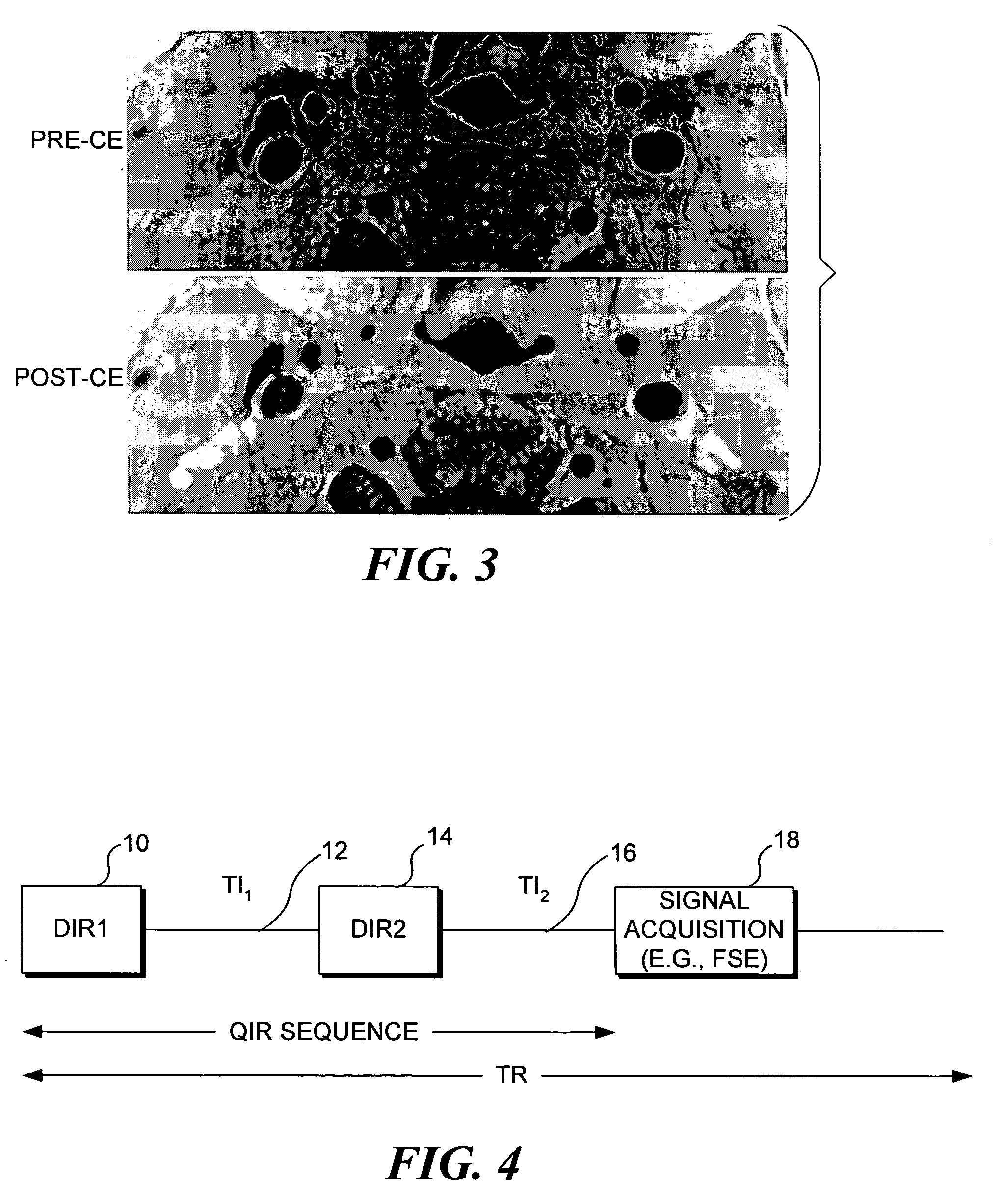
[0009]In addition, the black-blood image can be obtained at the site before infusion of the CE agent. Essentially, this image guarantee consistent suppression of the signal from blood using the same parameters that are used in the present invention for post-CE imaging. Because images before and after CE have been obtained using the identical method, any changes of signal intensity are caused only by the penetration of the CE agent into tissue. This information disclosed by the resulting images is used for the characterization of pathologic tissues in the cardiovascular system, such as atherosclerotic plaques, thrombi, myocardial infarction, and other conditions.
[0010]Another aspect of the invention is directed to a pulse sequence for quadruple inversion-recovery imaging. The pulse sequence includes two double inversion RF pulse pairs, each of which is followed by one of two inversion delays. Each double inversion pulse pair includes two inversion RF pulses, one of which is non-selective and the other of which is slice-selective or slab-selective. The pulse sequence also includes all RF and magnetic field gradient pulses needed to generate, encode, and acquire an MR signal. Optionally, the sequence may contain more than two double inversion blocks in order to improve blood suppression effect.

Problems solved by technology

However, DIR only provides efficient suppression of the blood signal within a narrow range of T1 values at any selected TI.

Method used


Image

  • Quadruple inversion recovery for quantitative contrast-enhanced black blood imaging
  • Quadruple inversion recovery for quantitative contrast-enhanced black blood imaging
  • Quadruple inversion recovery for quantitative contrast-enhanced black blood imaging

Examples


Embodiment Construction

Method

[0018]Sequence: In the present invention, suppression of the flowing blood signal and artifacts is achieved using a quadruple inversion recovery (QIR) pulse sequence. The QIR pulse sequence includes two double inversion pulse pairs, the first double inversion pulse pair being followed by an inversion delay period, TI1, and the second double inversion pulse pair being followed by an inversion delay period, TI2. The double inversion procedure is known from its use in a double inversion-recovery (DIR) pulse sequence. Use of the DIR sequence for achieving black-blood imaging is generally well known to those of ordinary skill in the art of MRI. It is also known that the DIR method provides efficient suppression of the blood signal in a narrow range of T1 values, which makes the use of DIR problematic in the presence of contrast enhancing agents, since such agents strongly affect the T1 of blood. However, in accord with the present invention, by performing a plurality of successive ...


PUM


Abstract

A contrast enhancement (CE) agent is infused into blood flowing through a site that is to be imaged with magnetic resonance imaging (MRI). Two double inversion procedures are carried out, forming a quadruple inversion recovery (QIR) pulse sequence. Each double inversion procedure comprises a non-selective and slice-selective inversion RF pulse. The first double inversion procedure is followed by a first predefined inversion delay period, TI1, and the second procedure by a second predefined inversion delay period, TI2. A black-blood image can thus be produced in which blood appears consistently black and tissues surrounding the blood, such as a vessel wall, heart, atherosclerotic plaque, or thrombus, are clearly visible. Unlike the prior art black-blood imaging technique, the QIR method does not require a precise knowledge of the T1 of the blood carrying the CE agent in order to suppress the signal and artifacts caused by the blood flowing through the site.

Description

RELATED APPLICATIONS[0001]This application is based on prior provisional patent application, Ser. No. 60 / 435,835, filed on Dec. 19, 2002, the benefit of the filing date of which is hereby claimed under 35 U.S.C. §119(e).GOVERNMENT RIGHTS[0002]This invention was made with U.S. Government support under Grant No. RO1-HL56874 awarded by NIH-NIHB1. The U.S. Government has certain rights in the invention.FIELD OF THE INVENTION[0003]This invention generally pertains to magnetic resonance imaging (MRI) and more specifically, pertans to a method, a pulse sequence, and a system that make use of a plurality of radiofrequency (RF) inversion pulses grouped into double-inversion procedures in order to suppress the signal from flowing blood having a variety of T1 relaxation times caused by the presence of a contrast-enhancing agent.BACKGROUND OF THE INVENTION[0004]Black-blood imaging is a technique developed to improve the visualization of blood vessels and the heart in magnetic resonance images s...

Claims


Application Information

Patent Timeline
IPC IPC(8): A61B5/05G01R33/28G01R33/563
CPCG01R33/5607G01R33/5601
Inventor YARNYKH, VASILY L.YUAN, CHUN
Owner UNIV OF WASHINGTON
Learn More