
Stain-directed molecular analysis for cancer prognosis and diagnosis

a molecular analysis and prognosis technology, applied in the field of stain-directed molecular analysis for cancer prognosis and diagnosis, can solve the problems of increased relative hazards of death for patients who delay obtaining a cancer consultation for at least two months, increased dna damage, and increased delay

Inactive Publication Date: 2010-02-09
DEN MAT HLDG
View PDF17 Cites 1 Cited by
  • Summary
  • Abstract
  • Description
  • Claims
  • Application Information

AI Technical Summary

Benefits of technology

"The patent describes a method for detecting cancer in human tissue by combining the benefits of two previous technologies: general screening and cellular molecular analysis. The method involves conducting a screening test to determine if there is a possibility of cancer in a specific region, applying a stain to the region to visually identify any cancerous or pre-cancerous tissue, and then analyzing the cells from the identified region using molecular analysis to determine if they show characteristics associated with cancer. This method can be used for any cells that can be visually inspected in vivo, including topical or internal tissues. The combination of steps provides a simple clinical protocol that can identify the locations of pre-cancerous or suspect sites well before they become visible."

Problems solved by technology

Patients who delay in obtaining a cancer consultation for at least two months have significantly higher relative hazards of death than do patients with a shorter delay.
However, these normally visible features and their characteristics may not be apparent until the tissue involved is advanced on the normal progression pathway of cancer.
It has been determined that a disruption or mutation in either the telomeric DNA or telomerase, or the intermediary RNA, can uncap the telomere, causing further damage to the DNA.
However, given the expanse of the human body's cellular tissue that could possibly propagate invasive cancer tissue, diagnostic techniques such as genetic, epigenetic, or mitochondrial molecular analysis are not effective early cancer detection methods, because the effectiveness of these techniques directly depend on obtaining tissue samples from the specific tissue sites containing cells which are propagating cancer.

Method used


Image

  • Stain-directed molecular analysis for cancer prognosis and diagnosis
  • Stain-directed molecular analysis for cancer prognosis and diagnosis
  • Stain-directed molecular analysis for cancer prognosis and diagnosis

Examples


working examples

[0059]The following examples illustrate for those skilled in the art a presently preferred embodiment of the invention and are not intended as a limitation of the scope thereof.

example i

Location of Suspect Tissue By Mashberg-type Clinical Protocol

Preparation of Clinical Test Solutions

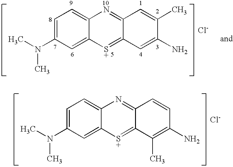
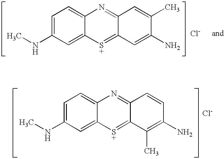
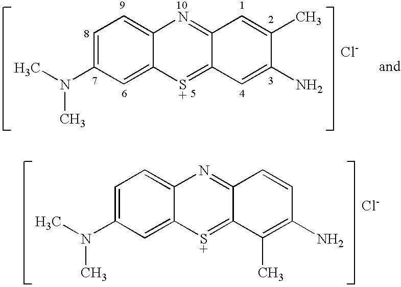
[0060]TBO (e.g., the product of Example I of U.S. Pat. No. 6,086,852), raspberry flavoring agent (IFF Raspberry IC563457), sodium acetate trihydrate buffering agent and H2O2 (30% USP) preservative (See U.S. Pat. No. 5,372,801), are dissolved in purified water (USP), glacial acetic acid and SD 18 ethyl alcohol, to produce a TBO test solution, having the composition indicated in Table A:

[0061]

TABLE AComponentWeight %TBO Product1.00Flavor.20Buffering Agent2.45Preservative.41Acetic Acid4.61Ethyl Alcohol7.48Water83.85100.00

[0062]Pre-rinse and post-rinse test solutions of 1 wt. % acetic acid in purified water, sodium benzoate preservative and raspberry flavor are prepared.

Clinical Protocol

[0063]The patient is draped with a bib to protect clothing. Expectoration is expected, so the patient is provided with a 10-oz. cup, which can be disposed of in an infectious waste container or the contents...

example ii

Genetic Alteration Molecular Analysis

[0073]58 samples of suspect tissue are obtained from various clinical sites practicing the screening procedure of Example 1. It is determined that genetic alteration analysis of two of these samples is not possible because there is inadequate material on the slides. In the remaining 56 cases neoplastic cells are carefully dissected (in cases with cancer) from normal tissue or epithelium (in all other cases) from normal tissue using a laser capture microdissection scope. This allows isolation of the cells and extraction of DNA for subsequent microsatellite analysis at three critical loci. In cases, there is insufficient DNA and further analysis is not possible. Two of the loci (D9S171 and D9S736) chosen for testing are on chromosomal region 9p21 which contains the p16 gene. A third marker (D3S1067) is located on chromosome 3p21. All molecular studies in the remaining 41 cases are done blinded without knowledge of the pathologic diagnosis.

[0074]Wit...


PUM

PropertyMeasurementUnit
structuresaaaaaaaaaa
HPLCaaaaaaaaaa
sizeaaaaaaaaaa

Abstract

The location at which tissue samples are obtained to determine whether cells exhibit characeristics associated with cell differentiation or cancer by molecular analysis is determined by topically applying to epithelial tissue a dye that selectively stains cancer and precancerous tissue.

Description

[0001]This application is a Continuation-In-Part of Then-Copending International Patent Application No. PCT / US00 / 26551, filed 26 Sep. 2000 (U.S. application Ser. No. 10 / 111,545, now abandoned), is a Division of U.S. patent application Ser. No. 10 / 017,007, filed Dec. 14, 2001, now abandoned) and a Continuation-In-Part of U.S. patent application Ser. No. 10 / 758,936, filed Jan. 14, 2004 now abandoned.[0002]This invention relates to a combined method for the early location and prognosis of tissue containing potentially invasive cancer cells, before the normal visual appearance of the tissue indicates potential development of invasive cancer, thus delaying a diagnosis of such tissue as precancerous or cancerous by conventional location, excision and histological procedures.[0003]In another respect, the invention relates to a combined method for location and detection of tissue containing such potentially invasive cancer cells, the normal visual appearance of which is anomalous, which may...

Claims


Application Information

Patent Timeline
Patent Type & Authority Patents(United States)
IPC IPC(8): C12Q1/68A61B10/00G01N33/48A61K49/00C12Q1/6886G01N1/30G01N33/574
CPCA61K49/006C12Q1/6886C12Q2600/156C12Q2600/118
Inventor BURKETT, DOUGLAS D.
Owner DEN MAT HLDG
Learn More