Method and apparatus for detection of vulnerable atherosclerotic plaque

Inactive Publication Date: 2009-10-13
BOARD OF RGT THE UNIV OF TEXAS SYST
View PDF72 Cites 200 Cited by
  • Summary
  • Abstract
  • Description
  • Claims
  • Application Information

AI Technical Summary

Benefits of technology

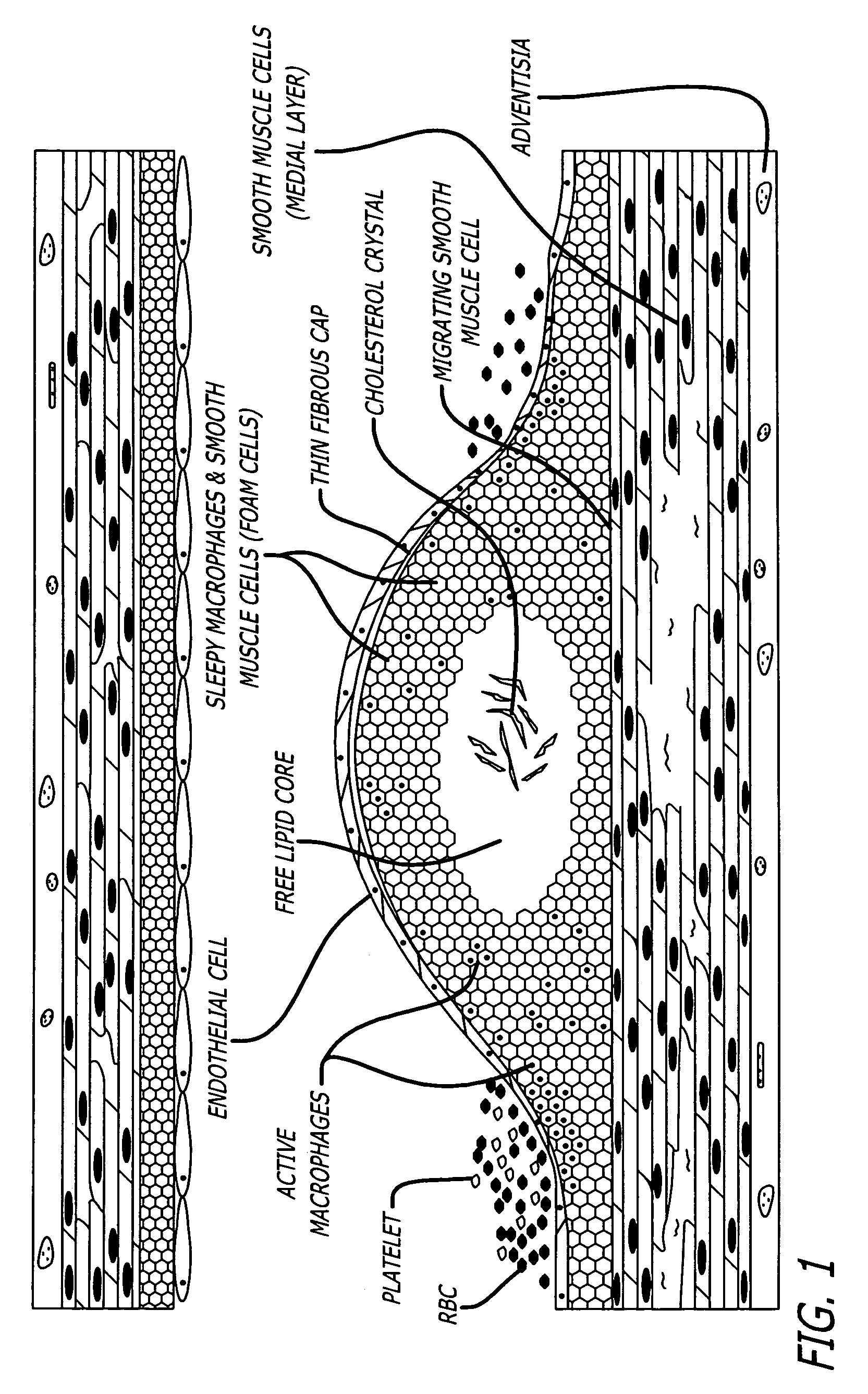
[0031]The methods and devices of the present invention provide an improvement over known methods and devices for distinguishing vulnerable plaque in a living vessel. As used herein, the term “at-risk”, “vulnerable,”“dangerous” or “unstable” plaque means an atherosclerotic plaque which, in a living vessel, is likely to develop a fissure, rupture or develop a thrombus leading to a life-threatening event. While most prior art methods are aimed at detecting and treating lumen stenosis, the present invention provides detection of an earlier stage of disease that precedes hemodynamically significant narrowing of the artery. This is an important advantage, especially since research over the past decade has established that these lesions account for most fatal myocardial infarctions (heart attacks) and many strokes. The methods and apparatus of the present invention provide for the first time a clinically useful tool to determine which lesion is dangerous and needs pre-emptive treatment.
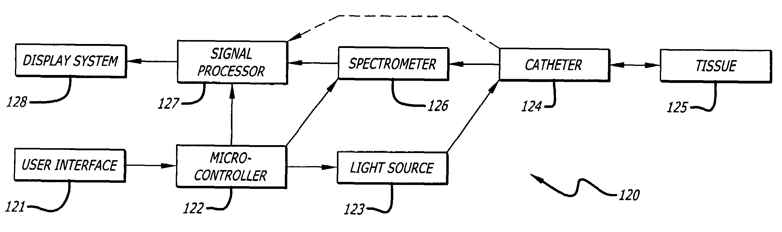
[0035]In accordance with the present invention, a method of detecting a vulnerable atherosclerotic plaque on a vessel wall is provided. The method includes simultaneously measuring in a site on a living vessel wall two or more chemical parameters associated with actively metabolizing cells (such as macrophages and other inflammatory cells, and smooth muscle cells). It is these highly active cells which are present in inflamed vulnerable atherosclerotic plaque. The qualitative or quantitative measurement of at least two of the following parameters is performed: pH, nitrosyl hemoglobin, nitrosyl tyrosine, glucose, lactate, oxidized collagen, oxyhemoglobin, reduced hemoglobin, oxidized cytochrome oxidase aa3. A preferred embodiment measures pH and oxidized collagen and, optionally, at least one other parameter from the following group: nitrosyl hemoglobin, nitrosyl tyrosine, glucose, lactate, oxyhemoglobin, deoxyhemoglobin and oxidized cytochrome oxidase aa3. In certain embodiments, the method also includes detecting leakage of dye into a plaque whereby a plaque cap fissure is revealed. The detection is accomplished by simultaneously measuring in a living vessel at least two chemical parameters, factors, or analytes, associated with inflamed vulnerable atherosclerotic plaque. This includes providing a fiber optic catheter having an illumination fiber bundle and a detection fiber bundle capable of, respectively, directing light onto or receiving light from a site on a vessel wall, the catheter having means for reducing optical interference by blood or other fluid within a vessel when undergoing examination. It also includes providing a source of 400-2500 nm wavelength light and a spectrometer, each operatively linked to the corresponding fiber bundle.
[0043]A method of treating a patient known to be suffering from atherosclerotic vascular disease and suspected of being at risk of experiencing a plaque rupture and / or an occlusive thrombotic event, is also provided by the present invention. The method comprises detecting an atherosclerotic plaque at risk of rupturing or thrombosing by taking chemical parameter measurements and dye sequestration measurements, as described above, and, optionally but preferably including monitoring the visible color image of the site and analyzing the visual image together with the chemical parameter and dye measurements, to obtain a predictive level of risk. Knowing the predicted level of risk for at least one site or group of sites on a wall of said vessel, at least one vulnerable plaque is then targeted for treatment. A localized therapeutic intervention or treatment is then administered to at least one of the targeted plaques. Suitable treatments can then be administered, for example, balloon angioplasty, laser angioplasty, heated balloon (RF, ultrasound or laser) angioplasty, surgical atherectomy, laser atherectomy, the placement of an appropriate stent, another conventional mechanical or irradiation treatment method, and pharmacological treatment regimens including anti-coagulants, fibrinolytic, thrombolytic, anti-inflammatory, anti-proliferative, immunosuppressant, collagen-inhibiting, endothelial cell growth-promoting, and conventional pharmacologically appropriate local treatments effective for reducing or eliminating inflamed plaque.
[0050]The methods and apparatus of the present invention go beyond the existing methods and devices for identifying a site of infection, cancer, a wound or the presence of autoimmune disease, and is particularly advantageous for identifying dangerous, rupture-prone plaque. Testing a vessel wall for combinations of specific chemical analytes associated with vulnerable plaque provides a better approach to distinguishing the particularly at-risk plaques from relatively stable atherosclerotic plaques in a living vessel.

Problems solved by technology

This defect is more pronounced once the organ increases its metabolic activity: this demands an even greater supply which cannot be satisfied due to the inappropriate perfusion resulting from the narrowing.
However, the “smart lasercatheter was disappointing in clinical trials (M. B. Leon, M. D., personal communication), and never became an accepted therapy.
These investigators noted the difficulty of defining and quantifying what is a “diseased state” of the tissue for obtaining meaningful comparative numbers.
Such a large blood clot may completely occlude the flow of blood through the artery, often causing injury to the heart or brain.
These are lesions that are hydronomically insignificant, as they cause no resistance to flow and can not be detected by any stress test.
A major diagnostic and prognostic dilemma for the cardiologist is how to predict which plaque is about to rupture.
Therefore, probing for lipid pools or even oxidized LDL would not be expected to provide an adequate way of diagnosing unstable plaque.
Each of these methods fails to recognize the functional activity level of atherosclerotic plaque however.
They do not focus on chemical factors indicative of activated macrophages, and fail to provide an adequate way to identify the truly dangerous plaques at risk of rupturing or thrombosing.
Methods relying on detection of calcium in vulnerable plaque are particularly problematic in view of the now widely-held view that calcified plaques are actually less likely to be vulnerable to rupture since the calcium deposit of the plaque can provide a type of physically supportive skeleton.
However, these techniques have not typically been used for monitoring inflammation.
Moreover, none of them have been utilized for diagnosing atherosclerotic plaque in a living vessel.
None of the known methods of measuring tissue oxygenation has previously been tested used to examine atherosclerotic plaque.
None of the known methods of measuring cytochrome aa3 has been applied to assessing the level of oxygenation in plaque.
This multi-parameter catheter is not suitable for measuring plaque oxygenation.
The prior art methods and devices fail to provide a clinically useful way of determining which lesion is dangerous and needs pre-emptive treatment.
Of the known methods directed at the detection of vulnerable plaque, none is adequate for distinguishing macrophage infiltrated, inflamed plaque from more stable plaque or normal vessel wall.
Moreover, none of the existing methods which attempt to locate the most dangerous plaques takes into account the considerable heterogeneity of macrophages in atherosclerotic plaque.
A major shortcoming of the prior methods is that none are capable of determining the presence of specific analytes and physiological variables correlated to macrophage activity.
Although near-infrared (NIR) spectroscopy has been employed to measure certain chemical factors, there is at the present time no method for studying analytes associated with vulnerable, rupture-prone atherosclerotic plaque.

Method used


Image

  • Method and apparatus for detection of vulnerable atherosclerotic plaque
  • Method and apparatus for detection of vulnerable atherosclerotic plaque
  • Method and apparatus for detection of vulnerable atherosclerotic plaque

Examples


Embodiment Construction

[0068]FIG. 1 depicts an atherosclerotic plaque within a vessel resulting from a chronic, continuous process of lipid infiltration into intima, endothelial injury, leukocyte migration into the subendothelial area, continuous lipid accumulation inside the macrophages, endothelial thickening and, smooth muscle cell proliferation and consequential increased matrix protein and collagen production and a fibrotic cap. In some (usually advanced) cases calcium deposition also occurs, leading to increased mass of the plaque, which causes it to gradually protrude eccentrically towards the endoluminal space and narrow the lumen of the artery. However, when a plaque instead progresses to a vulnerable plaque, as shown in FIG. 1, it is characterized largely by the presence of active macrophages, free lipid core, thin fibrous cap, endothelial denudation and microthrombi. Due to some not yet completely understood mechanism (perhaps hemodynamic parameters), disruption of these plaques usually occurs ...


PUM


Abstract

Methods for the detection of inflammation associated with vulnerable atherosclerotic plaque to prevent heart attack and stroke are disclosed. The methods are also applicable to detection of infection, cancer, wounds or auto-immune disease in the body. Certain embodiments of the new methods provide a way of predicting the level of vulnerability of an atherosclerotic plaque to rupture or thrombus formation by assessing via fiber optic NIR spectrophotometry the status of two or more parameters associated with inflamed atherosclerotic plaque in a vessel of a living patient. From these measurements such conditions as low pH, hypoxia, low glucose, oxidative stress or compounds abundant in vulnerable plaque such as oxidized LDL cholesterol and oxidized metabolites of NO, significant active macrophage population, thin plaque cap, as well as senescence and / or apoptosis of smooth muscle or endothelial cells are determined with the assistance of a suitably programmed microprocessor. By considering together the status of some or all of these conditions with respect to successive sites along a vessel wall, particular plaques which are at significant risk of rupturing or thrombosing can be distinguished from “normal” vessel wall and from “intermediate” and relatively stable or “lower risk” plaques. Sites having more of the indicator conditions would be considered most in need of prompt intervention, and certain combinations of parameter levels would be suggestive of relatively stable plaque.Also disclosed is a multi-parameter catheter and analytical processing assembly for use in the methods.

Description

CROSS REFERENCE TO A RELATED APPLICATION[0001]The present application is a continuation application of patent application Ser. No. 09 / 188,661, filed Nov. 9, 1998, and entitled “METHOD AND APPARATUS FOR DETECTION OF VULNERABLE ATHEROSCLEROTIC PLAQUE”.STATEMENT REGARDING FEDERALLY SPONSORED RESEARCH OR DEVELOPMENT[0002]This invention was made with government support under contact number DAMD17-98-18002 awarded by the United States Army. The government has certain rights in the invention.BACKGROUND OF THE INVENTION[0003]1. Field of the Invention[0004]The present invention generally relates to methods of diagnosing and treating inflammation, particularly inflamed atherosclerotic plaque at risk of rupture or thrombosis. More particularly, the methods of the invention pertain to the simultaneous detection of two or more chemical parameters associated with inflammation and / or active cellular metabolism in plaque. The invention also relates to multi-parameter catheter assemblies for irradia...

Claims


Application Information

Patent Timeline
IPC IPC(8): A61B5/00
CPCA61B5/01A61B5/6853A61B5/14539A61B5/1459A61B5/0507A61B1/04A61B1/044A61B1/046
Inventor CASSCELLS, III, S. WARDWILLERSON, JAMES T.NAGHAVI, MORTEZAGUO, BUJIN
Owner BOARD OF RGT THE UNIV OF TEXAS SYST
Why Eureka
Try Eureka