
Method to induce neovascular formation and tissue regeneration

a neovascular and tissue regeneration technology, applied in the field of neovascular formation and tissue regeneration, can solve the problems of ischemic heart, morbidity and mortality, many patients who are still symptomatic and cannot benefit from conventional therapy

Active Publication Date: 2009-07-21
FUNDACION UNIVRIA DR RENE G FAVALORO +1
View PDF4 Cites 3 Cited by
  • Summary
  • Abstract
  • Description
  • Claims
  • Application Information

AI Technical Summary

Benefits of technology

"The patent is about a method for using a protein called VEGF to promote the growth of new blood vessels and treat ischemic heart disease. The method involves administering VEGF through gene therapy, which involves introducing a gene that produces VEGF into the body. This approach has advantages over traditional protein therapy, as it allows for longer-term effects with lower doses. The patent also describes the use of VEGF to induce neovascular formation and tissue regeneration in mammals. Overall, the patent provides a novel and promising treatment for heart disease."

Problems solved by technology

Ischemic heart disease is the main cause of morbidity and mortality.
Despite recent advances in prevention and treatment of ischemic heart disease, there are many patients who are still symptomatic and cannot benefit from conventional therapy.
Therapeutic administration of VEGF is a significant challenge.
Furthermore, intravenous administration of high doses of VEGF protein is known to induce severe or refractory hypotension.
However, adenoviral gene therapy may induce inflammatory or immune reactions, especially after repeated doses.
These circumstances limit significantly the clinical use of this therapy.
Systemic administration of VEGF has been associated with undesired angiogenesis in peripheral tissues.
This condition results frequently in a significant loss of myocardial cells, reducing the contractile muscle mass.
However, this phenomenon occurs in a very small proportion of total cardiomyocyte population and under certain pathological conditions.
However, the efficiency of these methods in upper mammals has not been demonstrated yet.
Likewise, the failure of cardiomyocytes to replicate properly difficult adaptative hyperplasia (i.e. cell number increasing) as a response to other pathological conditions.
In most cases, cell adaptation is insufficient.
In consequence, the increased demands impair subendocardial perfusion even in the absence of coronary occlusion.
Finally, the combination of these factors leads to myocardial function detriment.

Method used


Image

  • Method to induce neovascular formation and tissue regeneration
  • Method to induce neovascular formation and tissue regeneration
  • Method to induce neovascular formation and tissue regeneration

Examples


example i

Induction of Ischemia

[0117]Eighty Landrace pigs weighing approximately 25 kg (approx. 3 months of age) were submitted to the following protocol: 1) each individual underwent clinical and laboratory assessment of good health; 2) a sterile thoracotomy was performed at the 4th left intercostal space under general anesthesia (induction: thiopental sodium 20 mg / kg; maintenance: 2% enflurane) and the circumflex coronary artery was dissected free from surrounding tissue at its proximal portion; 3) an Ameroid constrictor was placed embracing the origin of the circumflex coronary artery; and 4) the thoracotomy was repaired.

example ii

Basal Pre-Treatment Studies

[0118]Three weeks after the first surgery indicated in the previous example, basal (pre-treatment) studies were performed on the individuals. The studies were conducted under sedation with sufficient doses of intravenous sodium thiopental and under electrocardiographic control. Basal myocardial perfusion studies were performed on each individual. The left ventricular perfusion was quantified by single photon emission computed tomography (SPECT) utilizing an ADAC Vertex Dual Detector Camera System (ADAC Healthcare Information Systems Inc., USA). Sestamibi marked with 99mTc was utilized as contrast.

[0119]The studies were performed at rest and under pharmacological challenge with progressive doses of intravenous dobutamine. The dobutamine infusion was interrupted when heart rate was at least a 50% above the basal (rest) values.

[0120]Individuals fulfilling the inclusion criterium (hipoperfusion in a territory consistent with the circumflex coronary artery bed)...

example iii

Administration of VEGF Plasmid and Placebo Plasmid

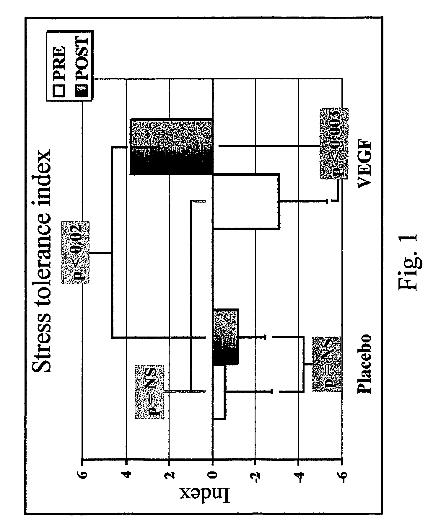
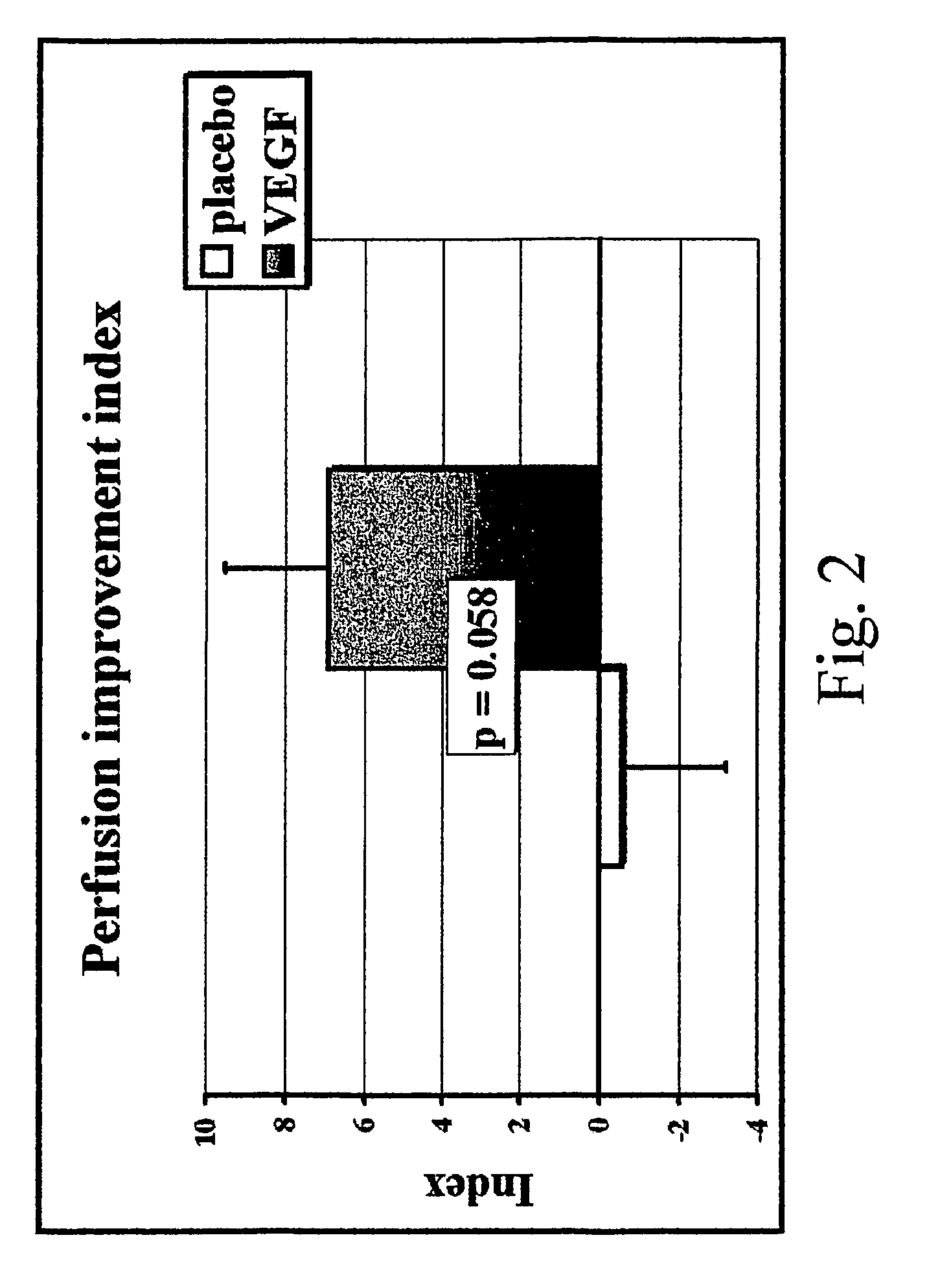
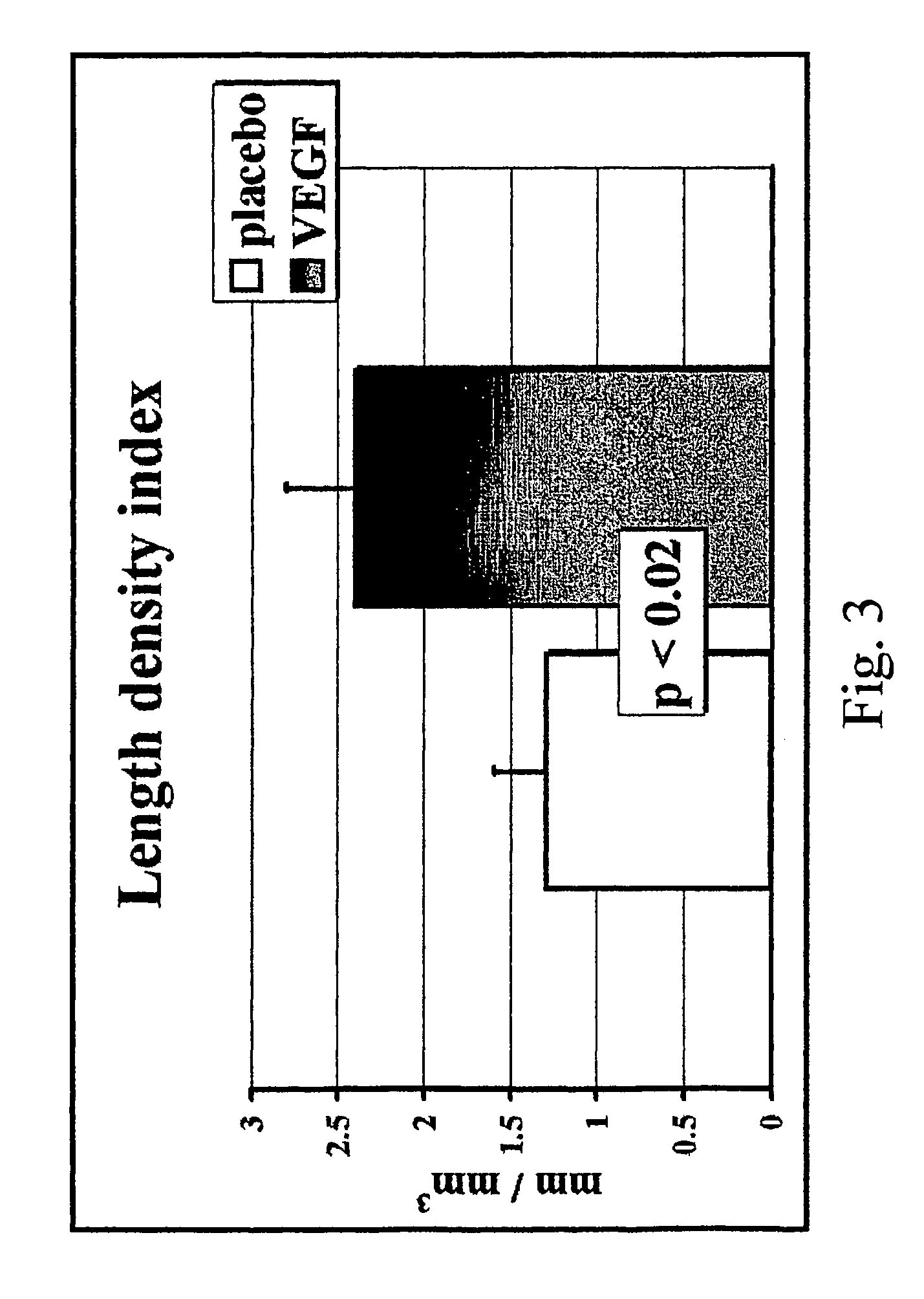
[0121]The twenty six individuals of the previous example were distributed in two groups: A first group consisting of 16 individuals (Group I) and a second group consisting of 10 individuals (Group II). Group I individuals were utilized to perform histopathological and physiological studies. Group II individuals were utilized to assess the presence and expression of the VEGF plasmid.

[0122]Group I individuals were randomized into two subgroups (Group I-T and Group I-P) with the same number of members (4 females and 4 males per subgroup). The treated group was designated Group I-T. The placebo group was designated Group I-P.

[0123]Group II individuals were randomized into two subgroups (Group II-T and Group II-P). Eight individuals were allocated to Group II-T. Two individuals were allocated to Group II-P. The treated group was designated Group II-T. The placebo group was designated Group II-P.

[0124]A sterile reopening of the previous th...


PUM

PropertyMeasurementUnit
angleaaaaaaaaaa
angleaaaaaaaaaa
concentrationaaaaaaaaaa

Abstract

The present invention relates, e.g., to a method for inducing arteriogenesis, lymphangiogenesis, vasculogenesis, or cardiomyogenesis, and / or for inducing mitosis or proliferation of a smooth muscle cell, a skeletal muscle cell, or a cardiomyocyte, comprising administering to a cell or tissue in need thereof a dose of a polynucleotide that encodes a vascular endothelial growth factor (VEGF), or that encodes a polypeptide comprising an active site of the VEGF. The coding sequence is operably linked to an expression control sequence; and the dose is sufficient to induce arteriogenesis, lymphangiogenesis, vasculogenesis, or cardiomyogenesis, and / or to induce mitosis or proliferation of a smooth muscle cell, a skeletal muscle cell, or a cardiomyocyte. In preferred embodiments of the invention, the method is a method of tissue regeneration, particularly of cardiomyogenesis; and the polynucleotide (or a polypeptide encoded by such a polynucleotide) is administered into the myocardium.

Description

[0001]This application is a continuation-in-part of PCT / US02 / 14508, filed May 13, 2002, which is incorporated by reference herein in its entirety.FIELD OF THE INVENTION[0002]This invention relates, e.g., to a method for inducing arteriogenesis, lymphangiogenesis, vasculogenesis, or cardiomyogenesis, or for inducing mitosis or proliferation of a smooth muscle cell, a skeletal muscle cell, or a cardiomyocyte, using a Vascular Endothelial Growth Factor (VEGF). For example, a gene therapy method is described for in vivo localized induction of neovascular formation or tissue regeneration in mammals utilizing VEGF.BACKGROUND OF THE INVENTION[0003]Ischemic heart disease is the main cause of morbidity and mortality. The epidemiological and socio-economical impact of coronary heart disease is remarkable. This disease causes million of deaths all over the world. See Murray, et al., Lancet, 349:1269-1276 (1997). In developed countries, it has been estimated that 5.3 million deaths attributable...

Claims


Application Information

Patent Timeline
Patent Type & Authority Patents(United States)
IPC IPC(8): A61K31/70A01N63/00A61K48/00C12N15/00C12N5/00A01N43/04A61KA61K6/00A61K9/127A61K31/7088A61K38/18A61K38/19C07H21/02C07H21/04C12N15/12C12N15/63C12N15/85C12P21/06
CPCA61K38/1866A61K48/0075A61K9/0019A61P9/00A61P9/10A61K9/127A61K48/0058
Inventor LAGUENS, RUBENARGUELLES, MARCELO L.VERA JANAVEL, GUSTAVO LEONIDASCROTTOGINI, JOSE ALBERTOMELO, CARLOS ALBERTOPICHEL, RICARDO HORACIOCRISCUOLO, MARCELO EDUARDO
Owner FUNDACION UNIVRIA DR RENE G FAVALORO
Learn More