Protein-based Streptococcus pneumoniae vaccines
a streptococcus pneumoniae and vaccine technology, applied in the field of protein-based streptococcus pneumoniae vaccines, can solve the problems of not overcoming the problem of coverage, and affecting the treatment effect of i>s. pneumoniae/i>
- Summary
- Abstract
- Description
- Claims
- Application Information
AI Technical Summary
Benefits of technology
Problems solved by technology
Method used
Image
Examples
example 1
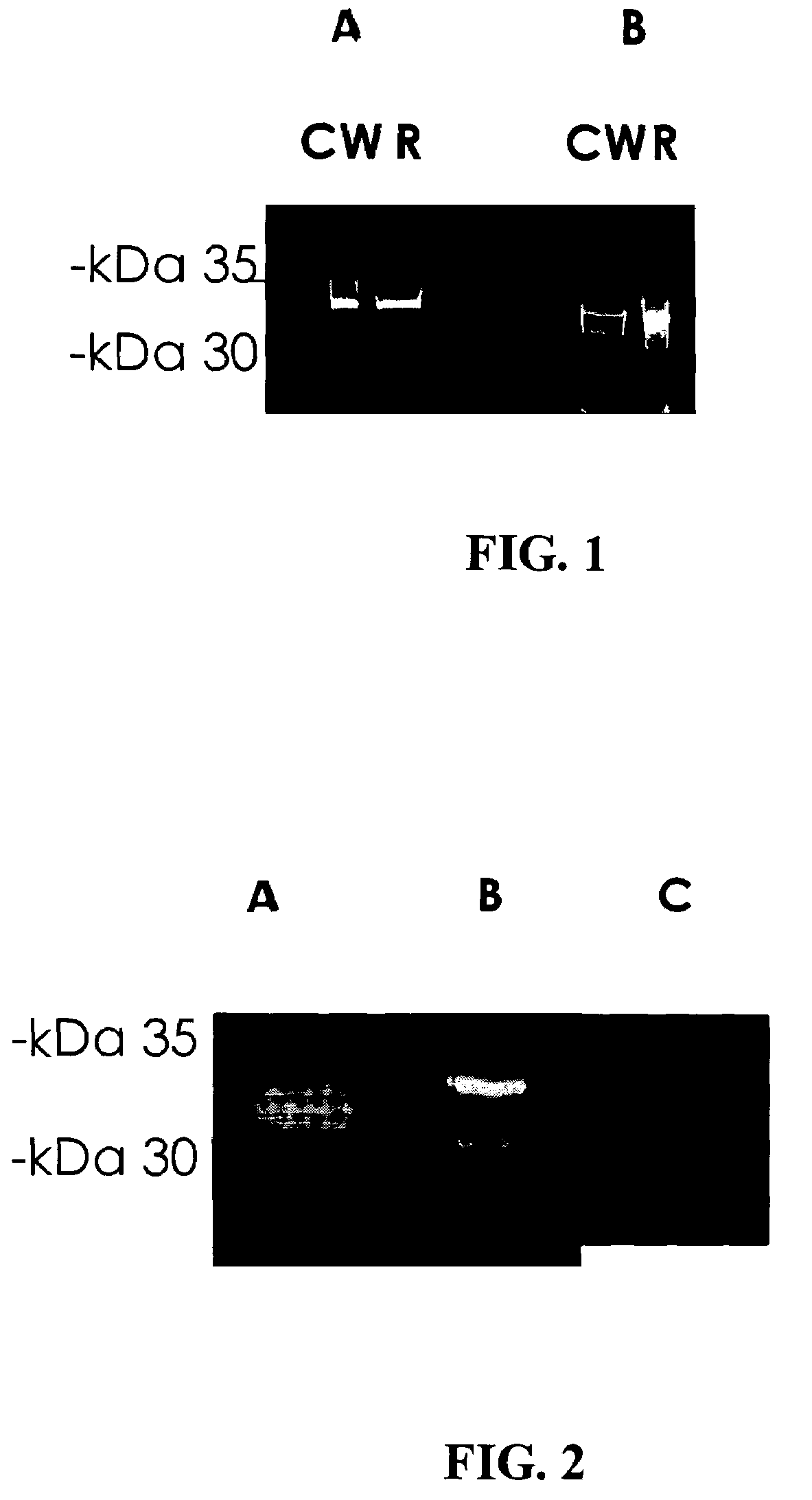
Prevention of S. pneumoniae Infection in Mice by Inoculation with S. pneumoniae Cell Wall Protein Fractions
Methods:
Bacterial Cells:
[0082]The bacterial strain used in this study was an S. pneumoniae serotype 3 strain. The bacteria were plated onto tryptic soy agar supplemented with 5% sheep erythrocytes and incubated for 17-18 hours at 37° C. under anaerobic conditions. The bacterial cells were then transferred to Todd-Hewitt broth supplemented with 0-5% yeast extract and grown to mid-late log phase. Bacteria were harvested and the pellets were stored at −70° C.
Purification of Cell Wall Proteins:
[0083]Bacterial pellets were resuspended in phosphate buffered saline (PBS). The resulting pellets were then treated with mutanolysin to release cell wall components. Supernatants containing the CW proteins were then harvested. Subsequently, the bacteria were sonicated and centrifuged and the resulting pellet containing the bacteria membranes (m) were lysed with 0.5% TRITON X-100 detergent.
Fr...
example 2
Determination of Age-related Immunoreactivity to S. pneumoniae Surface Proteins
[0086]The following study was carried out in order to investigate the age-related development of immunoreactivity to S. pneumoniae cell wall and cell membrane proteins.
[0087]Operating as described hereinabove in Example 1, a fraction containing cell wall proteins was obtained from a clinical isolate of S. pneumoniae. In addition, cell membrane proteins were recovered by solubilizing the membrane pellet (described hereinabove in Example 1) in 0.5% TRITON X-100 detergent. The cell wall and cell membrane proteins were separated by means of two-dimensional gel electrophoresis, wherein the proteins were separated using sodium dodecyl sulfate polyacrylamide gel electrophoresis (SDS-PAGE) in one dimension, and polyacrylamide gel isoelectric focusing in the other dimension.
[0088]The ability of serum prepared from blood samples of children aged 1.5, 2.5 and 3.5 years and adults to recognize the separated S. pneumo...
example 3
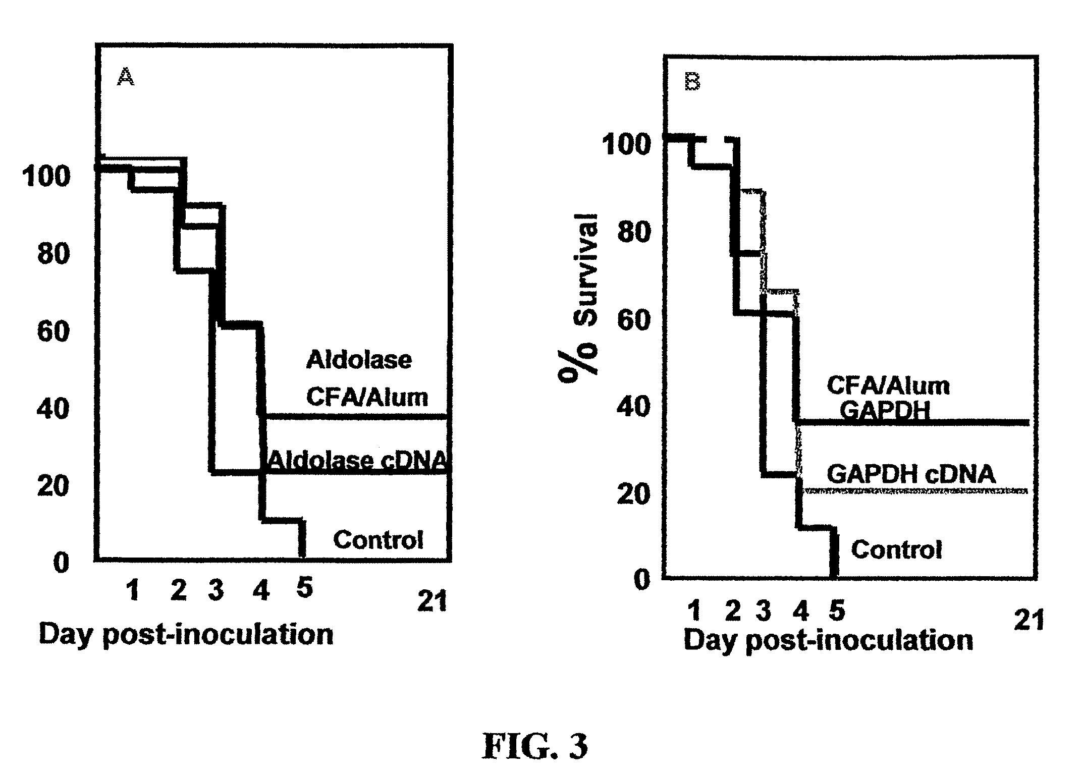
Prevention of S. pneumoniae Infection in Mice by Inoculation with Recombinantly-expressed S. pneumoniae Cell Surface Proteins
Cloning of Immunogenic S. pneumoniae Surface Proteins:
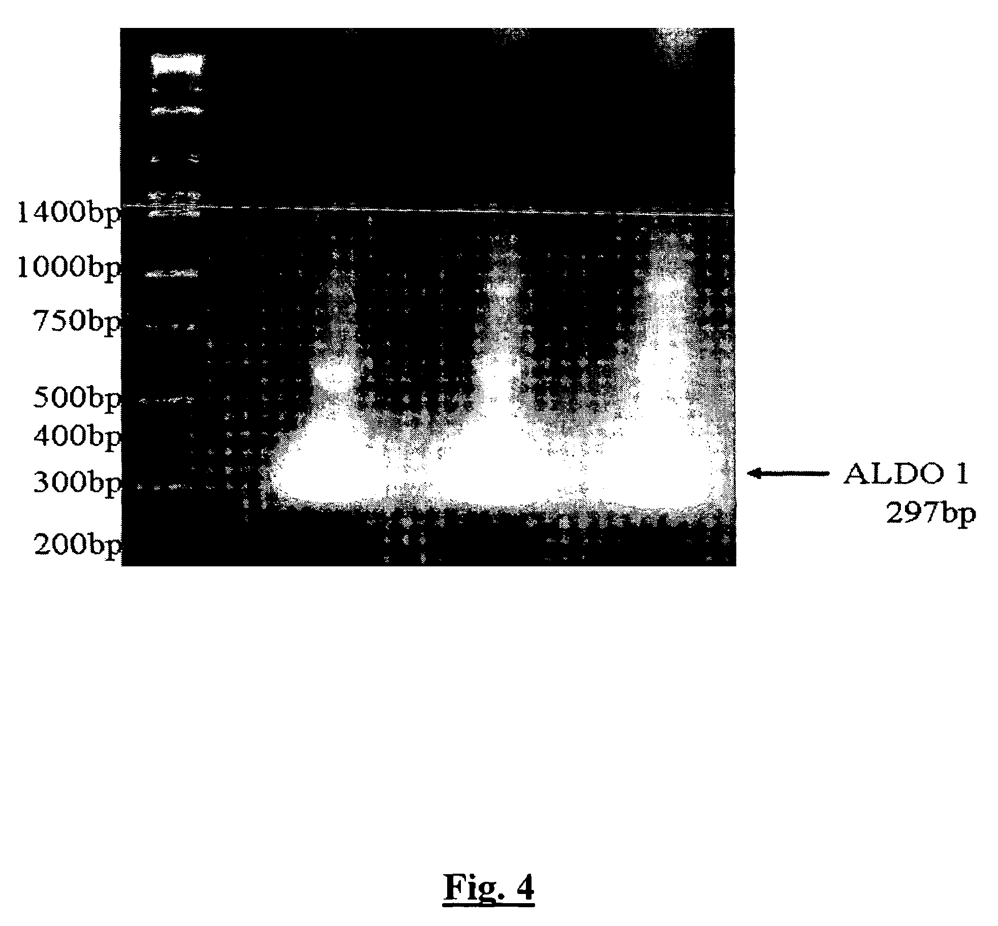
[0093]S. pneumoniae fructose-biphosphate aldolase (hereinafter referred to as “aldolase”) and GAPDH proteins were cloned into the pHAT expression vector (Clontech) and expressed in E. coli BL21 cells (Promega Corp., USA) using standard laboratory procedures. Following lysis of the BL21 cells, recombinant proteins were purified by the use of immobilized metal affinity chromatography (IMAC) on Ni—NTa columns (Qiagen) and eluted with imidazole. In a separate set of experiments, S. pneumoniae aldolase cDNAs were cloned into the pVAC expression vector (Invivogen), a DNA vaccine vector specifically designed to stimulate a humoral immune response by intramuscular injection. Antigenic proteins are targeted and anchored to the cell surface by cloning the gene of interest in frame between the IL2 signal sequence and ...
PUM
| Property | Measurement | Unit |
|---|---|---|
| length | aaaaa | aaaaa |
| chemical | aaaaa | aaaaa |
| weight | aaaaa | aaaaa |
Abstract
Description
Claims
Application Information
 - R&D
- Intellectual Property
- Life Sciences
- Materials
- Tech Scout
- Unparalleled Data Quality
- Higher Quality Content
- 60% Fewer Hallucinations
Browse by: Latest US Patents, China's latest patents, Technical Efficacy Thesaurus, Application Domain, Technology Topic, Popular Technical Reports.
© 2025 PatSnap. All rights reserved.Legal|Privacy policy|Modern Slavery Act Transparency Statement|Sitemap|About US| Contact US: help@patsnap.com


