
Automated brain MRI and CT prescriptions in Talairach space

a brain and talairach space technology, applied in the field of automatic brain mri and ct prescriptions in talairach space, can solve the problem that clinical diagnostic imaging still requires a significant amount of time to perform a detailed scan, and achieve the effect of minimizing intra- and inter-patient positioning variability

Inactive Publication Date: 2008-11-11
ABSIST +1
View PDF19 Cites 19 Cited by
  • Summary
  • Abstract
  • Description
  • Claims
  • Application Information

AI Technical Summary

Benefits of technology

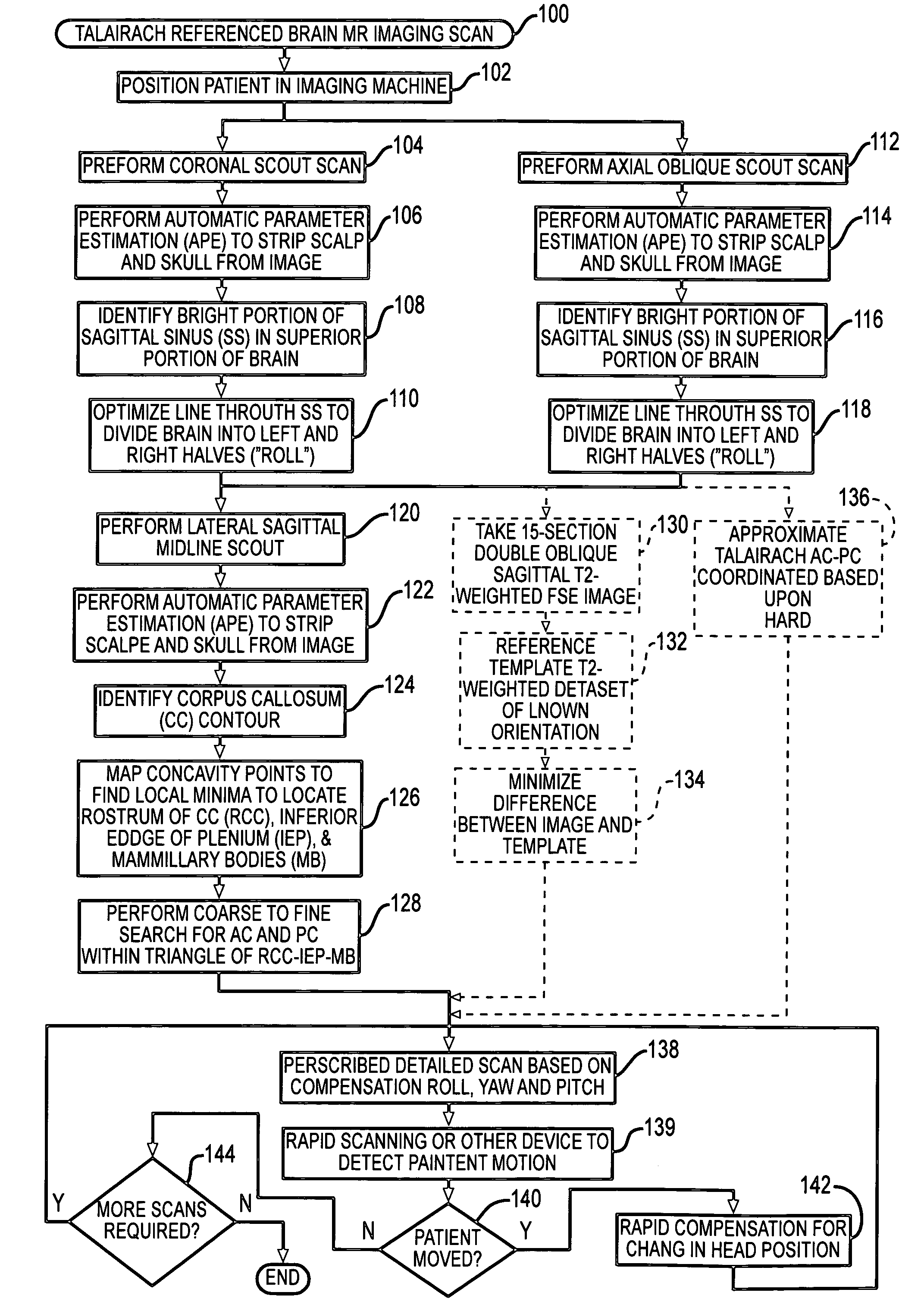
[0006]The invention overcomes the above-noted and other deficiencies of the prior art by providing an automated procedure that corrects for patient's head not being aligned by performing image analysis of a diagnostic image to determine coordinates of a Talairach anterior commissure (AC)—posterior commissure (PC) reference line. Thereby, subsequently prescribed scans are oriented, minimizing intra- and inter-patient positioning variability. Thus, further diagnostic and therapeutic measures may be employed that benefit from having known relative coordinates from scan to scan.
[0007]In one aspect of the invention, a method, medical diagnostic system, and computer product provide for identifying a head position of a patient undergoing diagnostic imaging by obtaining a diagnostic image of a patient's head. Image processing determines coordinates of the Talairach AC-PC reference line within the diagnostic image and defines a coordinate system of the diagnostic image with reference to the Talairach AC-PC reference line. Thereby, prescriptions for detailed scans and for checking for needed position corrections are enhanced.

Problems solved by technology

While advances in MRI and CT diagnostic systems have reduced the amount of time necessary, clinical diagnostic imaging still requires a significant amount of time to perform a detailed scan of a region of interest of a patient.
Once the detailed scan is obtained, an expert clinician must interpret the results, often challenged by inter-patient and intra-patient scan variations.

Method used


Image

  • Automated brain MRI and CT prescriptions in Talairach space
  • Automated brain MRI and CT prescriptions in Talairach space
  • Automated brain MRI and CT prescriptions in Talairach space

Examples


Embodiment Construction

[0021]BACKGROUND AND PURPOSE: Variability in patient head positioning may yield substantial inter-study image variance in the clinical setting. We describe and test three-step technologist and computer-automated algorithms designed to image the brain in a standard reference system and reduce variance.

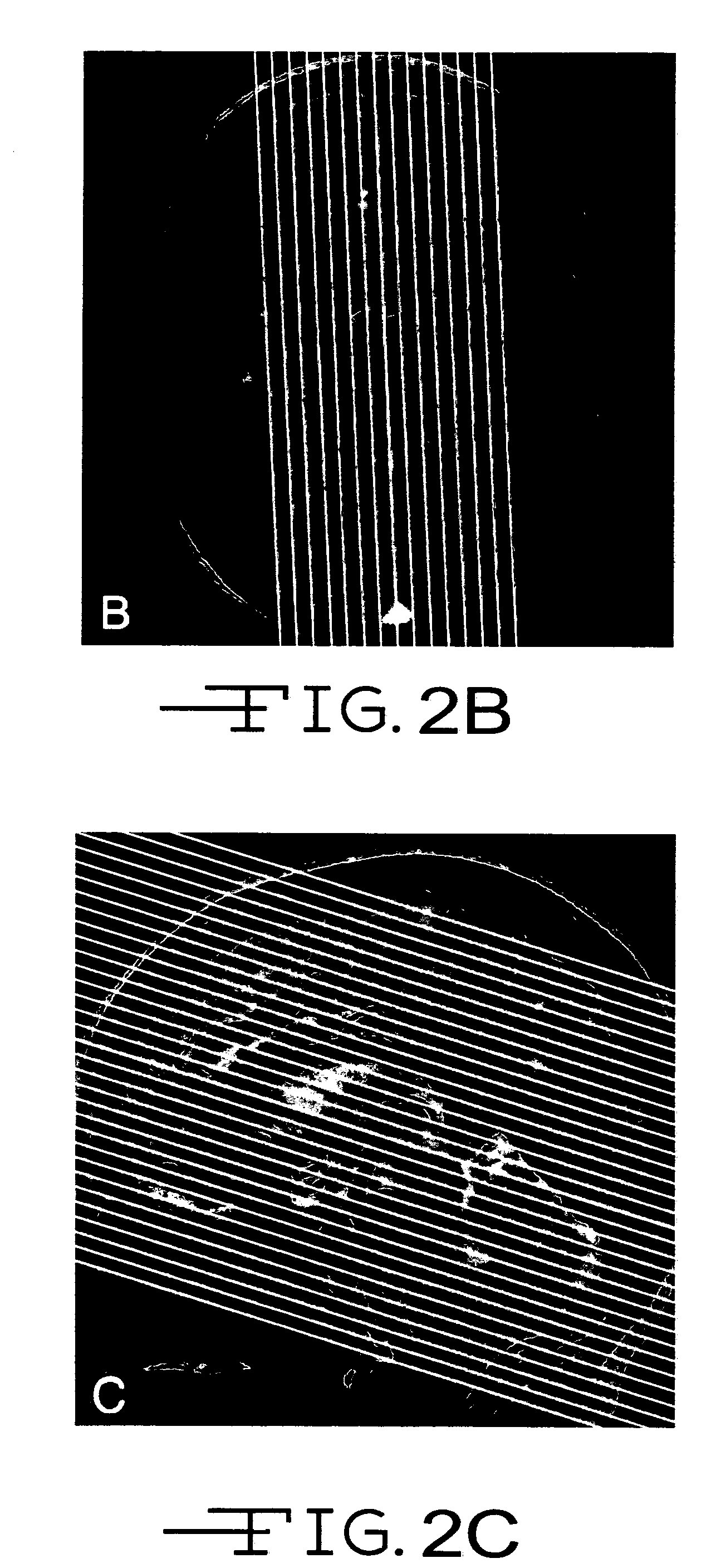
[0022]METHODS: Triple oblique axial images obtained parallel to the Talairach anterior commissure (AC)-posterior commissure (PC) plane were reviewed in a prospective analysis of 126 consecutive patients. Requisite roll, yaw, and pitch correction, as three authors determined independently and subsequently by consensus, were compared with the technologists' actual graphical prescriptions and those generated by a novel computer automated three-step (CATS) program. Automated pitch determinations generated with Statistical Parametric Mapping '99 (SPM'99) were also compared.

[0023]RESULTS: Requisite pitch correction (15.2°±10.2°) far exceeded that for roll (−0.6°±3.7°) and yaw (−0.9°±4.7°) in ...


PUM


Abstract

Brain Magnetic Resonance Imaging (MRI), Computerized Tomotography (CT), or other diagnostic modalities may employ a three-step procedure during initial (“scout”) cranial pre-scans that corrects for patient positioning (i.e., roll, yaw and pitch) to reduce inter- and intra-patient variation, thereby enhancing the diagnostic and comparative value of subsequent detail scans even across different diagnostic platforms. In MRI, for instance, locating the saggital sinus (SS) and optimizing a line to bisect the brain through this SS may be automated to correct for roll and yaw. By then identifying the contour of the corpus callosum in a lateral saggital scout scan, the Talairach anterior commissure (AC)—posterior commissure (PC) reference line may be found for correcting pitch. Prescription of detailed scans are improved, especially when the three-step correction is repeated periodically identifying the need to repeat a detailed scan or to adjust the coordinates of a subsequent scan.

Description

CROSS REFERENCE TO RELATED APPLICATIONS[0001]The present application hereby claims the benefit of the provisional patent application entitled “CLINICAL BRAIN MRI PRESCRIPTIONS IN TALAIRACH SPACE”, Ser. No. 60 / 455,969, filed on 18 Mar. 2003.FIELD OF THE INVENTION[0002]The present invention relates, in general, to devices that utilize a diagnostic imaging modality (e.g., magnetic resonance imaging (MRI) or x-ray-based Computerized Tomography (CT)), and more particularly to an automated system and method for interpreting a diagnostic screening scan to spatially orient to minimize inter- and intra-patient variability and to autoprescribe regions for a subsequent detailed scan.BACKGROUND OF THE INVENTION[0003]Medical diagnostic imaging is increasingly relied upon to detect various physical maladies. For example, X-ray-based Computerized Tomography (CT) is particularly suited for detection of skeletal injuries and dense cancerous lesions. As another example, Magnetic Resonance Imaging (MR...

Claims


Application Information

Patent Timeline
Patent Type & Authority Patents(United States)
IPC IPC(8): A61B5/05A61B6/03G01R33/56
CPCA61B6/032A61B6/488A61B6/501G01R33/5608Y10S128/922
Inventor WEISS, KENNETH L.
Owner ABSIST
Learn More