Membrane-based assays
a technology of membranes and assays, applied in the field of membrane-based assays, can solve the problems of time-consuming and inconvenient evaluation of candidate compounds affecting the properties of lipid bilayers, and is not well suited to evaluating this activity with any degree of detail
- Summary
- Abstract
- Description
- Claims
- Application Information
AI Technical Summary
Benefits of technology
Problems solved by technology
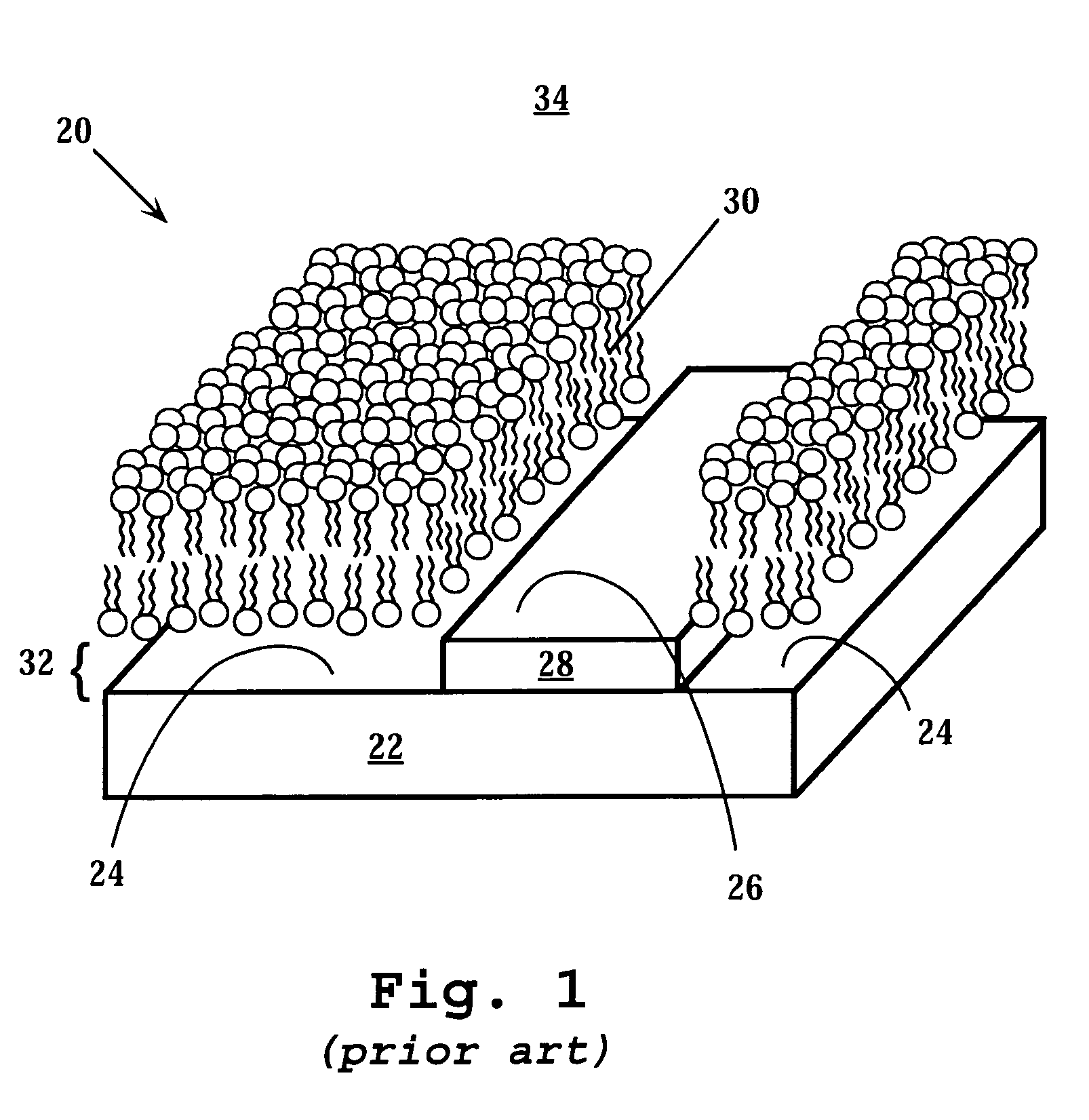
Method used
Image

Examples
example 1
Membrane Electrophoresis in a Surface Detector Device
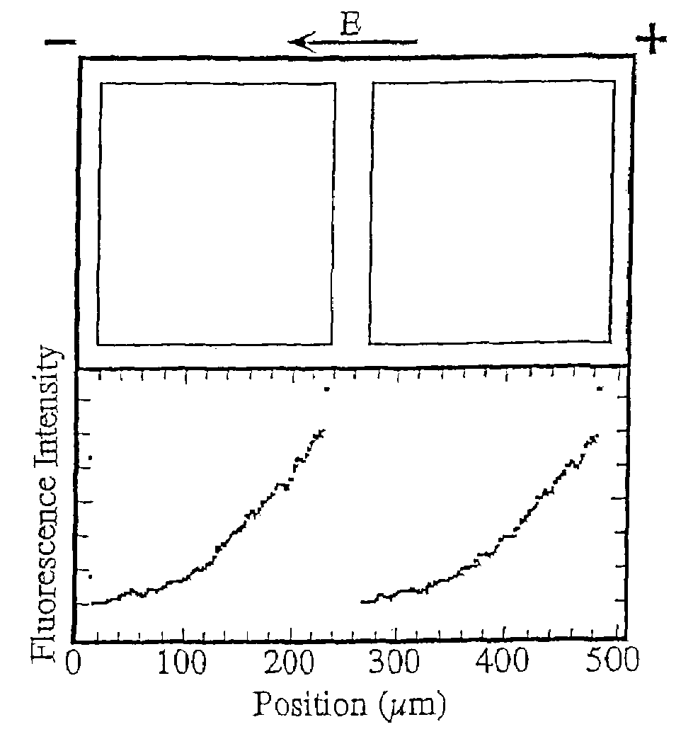
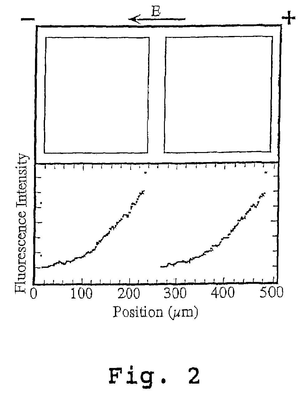
[0103]A surface detector device with 200 μm square corrals was prepared as described above and in Example 1 of U.S. Pat. No. 6,228,326 using L-α-phosphatidylcholine (PC) molecules doped with 1 mole percent of the fluorescently labeled lipid, Texas Red DHPE (Molecular Probes, Eugene, Oreg.).
[0104]Briefly, membranes were formed by contacting the patterned surface of the wafer support grids with a suspension, prepared as described above, containing ˜25 nm diameter unilamellar vesicles consisting primarily of molecules doped with 1 mole percent of the fluorescently labeled lipid, Texas Red DHPE. Excess vesicles were rinsed away while maintaining the membrane under the bulk aqueous solution at all times.
[0105]The fluidity of the supported bilayers on the bilayer-compatible surface regions was demonstrated by electrophoretic redistribution of charged membrane components. Electrophoresis was carried out using the technique described in M...
example 2
Screening Membrane Targets
[0108]The purpose of this experiment is to illustrate the feasibility of utilizing the surface detector array device, or “MembraneChip™,” for drug discovery, by utilizing the specific binding of the cholera toxin B subunits to the fluorescently-labeled glycolipid, ganglioside GM1. Any other membrane associated or integral components, such as other glycolipids, fatty acids, and sterols, can also be displayed on MembraneChips™ as membrane targets.
[0109]Cholera toxin is a membrane-targeting hexamer involving two different types of subunits, in an AB5 configuration. The toxin is secreted by Vibrio cholerae, a pathogen that accounts for over one million deaths, annually. The A subunit disrupts G-protein signaling, while the nontoxic B subunits are responsible for binding to cell surfaces. Each B subunit binds specifically to a pentasaccharide chain, that the ganglioside GM1 displays on its head region. GM1 is a naturally-occurring carbohydrate-rich sphingolipid ...
example 3
Detection of Binding by Alteration in Membrane Fluidity
[0115]Binding assays, such as those described above for cholera toxin, often require use of a labeled ligand to facilitate binding detection. Use of labeled ligands can create additional bottlenecks in screening processes, increase the expense of assays that require their use, or, depending on the specific label and its attachment point, alter ligand binding properties. Here we describe the use of membrane fluidity measurements to assay binding interactions. We illustrate that cholera toxin binding to membranes presenting GM1 decreases the lateral mobility of gangliosides within the membrane.
[0116]Materials and Methods
[0117]Cholera Toxin B subunit labeled with Alexa Fluor 594 was purchased from Molecular Probes (Eugene, Oreg.), and was received as 500 μg lyophilized B subunits. 0.25 ml water was added to create a 2.0 mg / ml solution, and 10 μl aliquots were partitioned and stored in a −20° C. freezer in a light-safe box.
[0118]Unl...
PUM
| Property | Measurement | Unit |
|---|---|---|
| bleached area | aaaaa | aaaaa |
| diffusion coefficients | aaaaa | aaaaa |
| thickness | aaaaa | aaaaa |
Abstract
Description
Claims
Application Information
 - R&D
- Intellectual Property
- Life Sciences
- Materials
- Tech Scout
- Unparalleled Data Quality
- Higher Quality Content
- 60% Fewer Hallucinations
Browse by: Latest US Patents, China's latest patents, Technical Efficacy Thesaurus, Application Domain, Technology Topic, Popular Technical Reports.
© 2025 PatSnap. All rights reserved.Legal|Privacy policy|Modern Slavery Act Transparency Statement|Sitemap|About US| Contact US: help@patsnap.com